SDRP Journal of Food Science & Technology (ISSN: 2472-6419)
DOI: 10.25177/JFST.4.2.RA.469
Research
Received Date: 24th Feb 2019 Copy rights: © This is an Open access article distributed Accepted Date: 17th Mar 2019 under the terms of International License. Published Date:24th Mar 2019
a Wine, Food and Molecular Biosciences Department, PO Box 85084, Lincoln University, Lincoln 7647, New Zealand
b College of Science, University of Canterbury, Private Bag 4800, Christchurch 8140, New Zealand
c Manaaki Whenua Landcare Research, PO Box 69040, Lincoln 7640, New Zealand
CORRESPONDENCE AUTHOR Ravi Gooneratne Tel. +64 3 423 0636 E-mail address: Ravi.Gooneratne@lincoln.ac.nz
CITATION
Ravi Gooneratne et al., Biochemical changes in sunflower plant exposed to silver nanoparticles / silver ions(2019)SDRP Journal of Food Science & Technology 4(2)
ABSTRACT摘要
When soil is contaminated with silver (Ag), plants take up Ag and is concentrated in roots and leaves, with its effects reflected in crop health and yield. This study investigated the toxicity of silver nanoparticles (AgNPs) and silver nitrate (AgNO3 as Ag+) tosunflowerseeds grown in soils amended with 150 mg/kg of Ag either as AgNPs or AgNO3. Exposure of the sunflower seeds to soils amended with Ag increased plant lipid peroxidation, activities of antioxidants enzymes (catalase, superoxide dismutase, glutathione-S-transferase), peroxidases (glutathione peroxidase pyrogallol peroxidase, guaiacol peroxidase), oxidases (ascorbate oxidase), urease, total phenolic compounds, vitamins (retinols, alpha-tocopherol and L-ascorbic acid) but inhibited chlorophyll, total carotenoids, total soluble carbohydrates, phenolic compounds and total soluble proteins. In general, AgNO3 increased the above-mentioned parameters in sunflower more than did AgNPs, except for the tested vitamins, which were more affected by AgNPs. The results showed that Ag accumulation in the root > leaf > stem and human food security risk is enhanced in sunflower seeds exposed to Ag compounds.
当土壤受到银(Ag)污染时,植物会吸收Ag并将其富集于根和叶中,其影响体现在作物健康状况和产量上。本研究考察了银纳米颗粒(AgNPs)和硝酸银(AgNO3,以Ag+形式)对生长于添加150 mg/kg Ag(以AgNPs或AgNO3形式)土壤中的向日葵种子的毒性。向日葵种子暴露于添加Ag的土壤后,植物脂质过氧化水平、抗氧化酶(过氧化氢酶、超氧化物歧化酶、谷胱甘肽-S-转移酶)、过氧化物酶(谷胱甘肽过氧化物酶、邻苯三酚过氧化物酶、愈创木酚过氧化物酶)、氧化酶(抗坏血酸氧化酶)、脲酶的活性以及总酚类化合物和维生素(视黄醇、α-生育酚和L-抗坏血酸)含量均升高,而叶绿素、总类胡萝卜素、总可溶性碳水化合物、酚类化合物和总可溶性蛋白则受到抑制。总体而言,除所测维生素受AgNPs影响更大外,AgNO3使向日葵上述各项指标升高的幅度均大于AgNPs。结果表明,Ag的累积量为根 > 叶 > 茎,且暴露于Ag化合物的向日葵种子会增加人类食品安全风险。
Keywords: Antioxidants, Chlorophyll, Silver ions, Silver nanoparticles, Sunflower, Total soluble protein, Silver toxicity, Oxidative stress.
关键词:抗氧化剂,叶绿素,银离子,银纳米颗粒,向日葵,总可溶性蛋白,银毒性,氧化胁迫。
1INTRODUCTION引言
Silver nanoparticles (AgNPs) are one of the most widely used industrial nanomaterials and are reported to be an efficient antimicrobial agent [1]. The special characteristics of AgNPs that have led to their largescale use in a variety of consumer products are also those which raise concern. Although sizes vary, by definition AgNPs are between 1 and 100 nm in three dimensions, with positive or negative charge depending on their coating material [2]. In general, nanoparticles (NPs) have a higher surface-to-volume ratio, which increases their reactivity with biological fluids [3]. The increased use of AgNPs in different industries may cause toxic effects on the environment because of improper discharge of wastes, and they may end up as contaminants in soil when sewage sludge is used as a fertilizer, with uncertain effects on plant crops and may cause soils to become infertile [4, 5].
银纳米颗粒(AgNPs)是应用最广泛的工业纳米材料之一,据报道是一种高效的抗菌剂[1]。AgNPs的特殊性质使其被大规模应用于多种消费品中,而这些性质恰恰也引发了人们的担忧。尽管尺寸不一,但按定义,AgNPs在三个维度上的尺寸均介于1~100 nm之间,并根据其包覆材料带正电荷或负电荷[2]。一般而言,纳米颗粒(NPs)具有较高的比表面积(表面积与体积之比),从而增强了其与生物体液的反应活性[3]。由于废弃物排放不当,AgNPs在各行业中的日益广泛使用可能对环境产生毒性效应;当污水污泥被用作肥料时,AgNPs最终可能成为土壤污染物,对农作物产生不确定的影响,并可能导致土壤肥力丧失[4, 5]。
The sunflower (Helianthus annuus L.; Asteraceae) is an herbaceous annual, grown commercially for its edible oil-bearing seeds. Each flower head produces hundreds of grey-white conical-shaped seeds, which are an excellent source of health benefitting nutrients like water- and fat-soluble vitamins. Each 100 g of seeds contains c. 35.17 g of alpha-tocopherol, essential macro-elements such as magnesium (Mg) and phosphorus (P), and micro-minerals such as copper (Cu), manganese (Mn), and selenium (Se). Packed with essential polyunsaturated fatty acids (linoleic acid, oleic acid), sunflower seeds are also a good source of proteins and amino acids that are essential for growth. Because sunflower plants are to some extent able to extract toxic elements from soil, they have been planted in contaminated soils to clean the soil from toxic elements [6]. However, exposure of sunflowers to AgNPs/Ag+ is known to cause oxidative stress and create reactive oxygen species (ROS), and as a result the activity levels of antioxidant defence enzymes and lipid peroxidation (LPO) increase.
向日葵(Helianthus annuus L.;菊科)是一种一年生草本植物,因其可食用的含油种子而被商业化种植。每个花盘可产生数百粒灰白色圆锥形种子,这些种子是水溶性和脂溶性维生素等有益健康营养素的优质来源。每100 g种子约含35.17 g α-生育酚、镁(Mg)和磷(P)等必需常量元素,以及铜(Cu)、锰(Mn)和硒(Se)等微量矿物质。向日葵种子富含必需多不饱和脂肪酸(亚油酸、油酸),同时也是生长所必需的蛋白质和氨基酸的良好来源。由于向日葵在一定程度上能够从土壤中提取有毒元素,因此已被种植于污染土壤中以清除土壤中的有毒元素[6]。然而,已知向日葵暴露于AgNPs/Ag+会引起氧化胁迫并产生活性氧(ROS),进而导致抗氧化防御酶活性和脂质过氧化(LPO)水平升高。
Studies have looked at the effect of other heavy metals on sunflower and an excess of Fe, Cu or Cd has been found to produce oxidative damage in sunflower leaves [7]. Treatment of sunflower with 0.5 mM iron (Fe2+), Cu2+ or cadmium (Cd2+) for 12 h increased
已有研究考察了其他重金属对向日葵的影响,发现过量的Fe、Cu或Cd会对向日葵叶片造成氧化损伤[7]。用0.5 mM铁(Fe2+)、Cu2+或镉(Cd2+)处理向日葵12 h后,
LPO and lipoxygenases when exposed to sunlight, but decreased chlorophyll A and B and glutathione (GSH) concentrations by 30%, 40%, and 15% respectively [7]. Free radical scavengers such as sodium benzoate or mannitol protected many of these enzymes from inactivation, but not superoxide dismutase (SOD). Exposure of sunflower to Fe2+ and Cd2+ ions decreased SOD while the Cu2+ ions increased SOD. However, all these metals increased a variety of other antioxidant enzymes such as catalase (CAT), ascorbate peroxidase, glutathione reductase and dehydroascorbate reductase.
在日光照射下LPO和脂氧合酶水平升高,而叶绿素A、叶绿素B和谷胱甘肽(GSH)浓度分别降低了30%、40%和15%[7]。苯甲酸钠或甘露醇等自由基清除剂可保护其中许多酶免于失活,但对超氧化物歧化酶(SOD)无保护作用。向日葵暴露于Fe2+和Cd2+离子后SOD降低,而暴露于Cu2+离子后SOD升高。然而,所有这些金属均使过氧化氢酶(CAT)、抗坏血酸过氧化物酶、谷胱甘肽还原酶和脱氢抗坏血酸还原酶等多种其他抗氧化酶升高。
Sunflower plants exhibit a strong potential to form Ag+from AgNPs compared to Basella alba (Basellaccea), Oryza sativa, Saccharum officinarum, Sorghum bicolor and Zea mays (Poaceae) [8]. Sewalem et al. 2014 [9], studying the role of sunflower as a potential phytoremediator of heavy metals, found that 88.8% of Cd accumulated in roots, inducing a low level of LPO, membrane leakage and poor root growth, whereas 71.4% of Pb was translocated to the shoots and hence affected photosynthesis.
与Basella alba(落葵科)、Oryza sativa, Saccharum officinarum, Sorghum bicolor和Zea mays(禾本科)相比,向日葵表现出较强的由AgNPs生成Ag+的潜力[8]。Sewalem et al. 2014 [9]在研究向日葵作为重金属潜在植物修复植物的作用时发现,88.8%的Cd累积于根部,引起较低水平的LPO、膜渗漏和根系生长不良;而71.4%的Pb转运至地上部,从而影响了光合作用。
Silver nanoparticles at concentrations of 20, 40, 60, 80 and 100 mg kg-1 impacted on plant growth parameters such as leaf surface area, chlorophyll, and carbohydrate, all of which affected growth of the common bean (Phaseolus vulgaris) and corn (Zea mays), promoting plant growth at low doses and inhibition at high concentrations [10].
浓度为20、40、60、80和100 mg kg-1的银纳米颗粒影响了叶面积、叶绿素和碳水化合物等植物生长参数,这些参数均影响菜豆(Phaseolus vulgaris)和玉米(Zea mays)的生长,表现为低剂量促进植物生长、高浓度抑制植物生长[10]。
Exposure of Bacopa monnieri to Ag+ and AgNPs enhanced peroxidase and catalase activity [11]. Qian and colleagues [12] studied the effect of Ag on the transcription of antioxidant and aquaporin genes, and the balance between antioxidant and oxidant status, and concluded that AgNPs were more toxic than Ag+ (in AgNO3). When watermelon plants were exposed to 0, 30, 60, 90 and 200 mg L-1 of Ag+applied at intervals of 8 days throughout the crop cycle, a dose responsive increase in Ag concentration occurred in both roots and shoots and the amount of antioxidant in fruits increased by 3-fold in those exposed to 30 mg L-1 Ag+but the lycopene concentration declined [13].
Bacopa monnieri暴露于Ag+和AgNPs后,过氧化物酶和过氧化氢酶活性增强[11]。Qian等[12]研究了Ag对抗氧化基因和水通道蛋白基因转录以及抗氧化与氧化状态平衡的影响,得出AgNPs的毒性大于Ag+(AgNO3中)的结论。在整个作物生长周期内每隔8天对西瓜植株施加0、30、60、90和200 mg L-1的Ag+,结果根和地上部的Ag浓度均呈剂量依赖性升高;暴露于30 mg L-1 Ag+的植株果实中抗氧化物含量增加了3倍,但番茄红素浓度下降[13]。
This study therefore aimed to investigate the effects of AgNPs and Ag+ (as AgNO3) on the sunflower plant and compared their effect on the activities of antioxidant defence enzymes [SOD, CAT, glutathione peroxidase (GPx), glutathione-S-transferase (GST)], other peroxidases (guaiacol peroxidase, pyrogallol peroxidase), ascorbate oxidase, urease, and LPO, antioxidant vitamins (A, E and C), chlorophyll A and B, total carotenoids, carbohydrate and protein concentration.
因此,本研究旨在考察AgNPs和Ag+(以AgNO3形式)对向日葵植株的影响,并比较二者对以下指标的作用:抗氧化防御酶[SOD、CAT、谷胱甘肽过氧化物酶(GPx)、谷胱甘肽-S-转移酶(GST)]、其他过氧化物酶(愈创木酚过氧化物酶、邻苯三酚过氧化物酶)、抗坏血酸氧化酶、脲酶的活性和LPO,抗氧化维生素(A、E和C),叶绿素A和B,总类胡萝卜素,以及碳水化合物和蛋白质浓度。
2MATERIALS AND METHODS材料与方法
2.1. Silver nanoparticles synthesis and quantification2.1. 银纳米颗粒的合成与定量
AgNPs were freshly prepared on the day of the experiments. Citrate-coated AgNPs were synthesized from AgNO3, reduced by ferrous sulphate hepta-hydrate, and coated with tri-sodium citrate dihydrate [14]. This method produced spherical-shaped particles, which did not aggregate due to the electrostatic repulsion of the citrate coating agent.
AgNPs于试验当天新鲜制备。以AgNO3为原料,用七水合硫酸亚铁还原,并以二水合柠檬酸三钠包覆,合成柠檬酸包覆的AgNPs[14]。该方法制得球形颗粒,由于柠檬酸包覆剂的静电排斥作用,颗粒不发生团聚。
The Ag concentration in AgNPs was determined by flame atomic absorption spectroscopy (FAAS) (Model A.6200, Shimadzu, Tokyo, Japan), calibrated with the Merck ICP certiPUR multi-element standard (Sigma-Aldrich, Castle Hill, New South Wales).
采用火焰原子吸收光谱法(FAAS)(Model A.6200, Shimadzu, Tokyo, Japan)测定AgNPs中的Ag浓度,并以Merck ICP certiPUR多元素标准溶液(Sigma-Aldrich, Castle Hill, New South Wales)进行校准。
2.2. Charge measurement and particle size2.2. 电荷测定与粒径
The average mean particle size and zeta potential of AgNPs were determined using a Zetasizer Nano ZS (Malvern Instruments, Worcestershire, UK) at 25°C. The conditions employed were: He/Ne laser (wavelength = 633 nm), scattering angle 90°, refractive index 1.33, and viscosity 0.887 mPa. Prior to the measurements, the concentration of the AgNPs was diluted 200-fold with water.
采用Zetasizer Nano ZS(Malvern Instruments, Worcestershire, UK)于25°C下测定AgNPs的平均粒径和Zeta电位。测定条件为:He/Ne激光(波长 = 633 nm),散射角90°,折射率1.33,黏度0.887 mPa。测定前,将AgNPs用水稀释200倍。
Silver nanoparticle morphology and size were determined using Transmission Electron Microscopy (TEM). Silver nanoparticles were diluted with deionized water to obtain a more uniform distribution and transferred directly onto a TEM gold-carbon grid for size characterization, using a high-resolution TEM (Philips CM200, Amsterdam) fitted with a Gatan digital camera.
采用透射电子显微镜(TEM)测定银纳米颗粒的形貌和粒径。将银纳米颗粒用去离子水稀释以获得更均匀的分布,然后直接转移至TEM金-碳载网上,使用配备Gatan数码相机的高分辨率TEM(Philips CM200, Amsterdam)进行粒径表征。
2.3. Soil amendment and plant sampling/ preparation2.3. 土壤添加处理与植物采样/制备
Templeton Silt Loam (TSL) soil (Table 1) (c. 100 kg) was collected from the Johnson Memorial Laboratory grounds, Lincoln University, Canterbury, New Zealand (43°38'44.67"S, 172°27'3.19"E), sieved and dried. The soil was divided into three portions: one portion was mixed with 150 mg/kg of Ag as AgNPs, the second with an equivalent amount of Ag as Ag- NO3. (This dose was chosen based on a study by N. Saleeb, which found that Ag at 200 mg/kg was mildly toxic to ryegrass) and the third portion was not treated (control). Four 8-L pots per treatment (control, AgNPs, AgNO3; n = 12) were each filled with 5 kg of soil from their respective portion (c. 20 kg per treatment).
Templeton粉砂壤土(TSL)(表1)(约100 kg)采自新西兰坎特伯雷林肯大学Johnson Memorial Laboratory场地(43°38'44.67"S, 172°27'3.19"E),经过筛和干燥处理。将土壤分为三份:第一份混入150 mg/kg的Ag(以AgNPs形式),第二份混入等量的Ag(以AgNO3形式)(该剂量的选择依据N. Saleeb的一项研究,该研究发现200 mg/kg的Ag对黑麦草具有轻度毒性),第三份不作处理(对照)。每个处理(对照、AgNPs、AgNO3;n = 12)设4个8 L盆,每盆装入相应部分的土壤5 kg(每个处理约20 kg)。
| Parameter参数 | Mean (SEM)均值(SEM) |
|---|---|
| Sand/silt/clay (%) | 76/20/4 |
| pH (H2O) | 5.1 |
| CEC (meq/100 g) | 12.3 |
| C (%) | 3.3 (0.03) |
| N (%) | 0.3 (0.00) |
| P (mg/kg) | 732 (11) |
| S (mg/kg) | 383 (6) |
| Ca (mg/kg) | 3329 (58) |
| Mg (mg/kg) | 3426 (71) |
| K (mg/kg) | 2541 (279) |
| Cd (mg/kg) | 0.13 (0.00) |
| Zn (mg/kg) | 70 (2) |
| Cu (mg/kg) | 5 (0) |
| B (mg/kg) | 7.3 (1.0) |
| Fe (mg/kg) | 17727 (353) |
| Mn (mg/kg) | 357 (20) |
| Ag (mg/kg) | <2 |
Three sunflower seeds (Helianthus annus, family Asteracear) were planted in each pot then water added to field capacity. Pots were watered daily to field capacity. After 2 weeks, two sunflower plants were
每盆播种3粒向日葵(Helianthus annus,Asteracear科)种子,然后浇水至田间持水量。每天浇水至田间持水量。2周后,每盆拔除两株向日葵
removed from each pot, leaving one per pot. The plants were harvested after 53 days, the roots separated, and the stem divided into four equal sections (labelled from the base to the top of the plant; S1, S2, S3, S4). All leaves from each plant were collected along with the flower, and seeds. The different sets of plant parts were then allocated to two groups. One half of each set of plant parts was kept in plastic bags and stored at -200C for measurement of LPO, total soluble protein, total phenolic compounds (TPC), total soluble carbohydrates, chlorophyll A and B, total carotenoids, antioxidant vitamins A, E and C, and activities of antioxidant enzymes, urease, and peroxidases (POX). The frozen sunflower leaves were homogenized with liquid nitrogen (N2) and the powder stored at −20°C until analysis. Leaf material (1 g) in liquid N2 was homogenized with 3 ml of 0.1 M phosphate buffer (pH 6.8) using a homogenizer (YunlinLi FJ-200, Shanghai, China) in ice and centrifuged (Beckman J2-MI, USA) at 16,000 rpm (30,390g RCF) for 20 min at 4°C and the supernatant collected for antioxidant enzyme analysis. All the parameters were measured using spectrophotometry (Shimadzu UV-VIS 1280, Tokyo, Japan). The other half was dried at 65°C for 72 h and then powdered in a tissue analyzer (Tissuelyser II Qiagen, USA). The powdered plant parts were subjected to acid (a mixture of 3 parts of HNO3 and I of HCl acids) digestion to determine the Ag concentration using inductive coupled plasma-optical emission (ICP-OES).
植株,每盆保留一株。播种53天后收获植株,分离根系,并将茎等分为四段(自植株基部至顶端依次标记为S1、S2、S3、S4)。收集每株植物的全部叶片以及花和种子。随后将各组植物部位分为两份。每组植物部位的一半装入塑料袋,于-200C下保存,用于测定LPO、总可溶性蛋白、总酚类化合物(TPC)、总可溶性碳水化合物、叶绿素A和B、总类胡萝卜素、抗氧化维生素A、E和C,以及抗氧化酶、脲酶和过氧化物酶(POX)的活性。冷冻的向日葵叶片在液氮(N2)中研磨匀浆,粉末于−20°C下保存直至分析。取液氮中的叶片材料(1 g),加入3 ml 0.1 M磷酸盐缓冲液(pH 6.8),在冰浴中用匀浆机(YunlinLi FJ-200, Shanghai, China)匀浆,于4°C下以16,000 rpm(30,390g RCF)离心(Beckman J2-MI, USA)20 min,收集上清液用于抗氧化酶分析。所有参数均采用分光光度法(Shimadzu UV-VIS 1280, Tokyo, Japan)测定。另一半于65°C下烘干72 h,然后用组织研磨仪(Tissuelyser II Qiagen, USA)粉碎。粉碎后的植物样品经酸(3份HNO3与1份HCl的混合酸)消解,采用电感耦合等离子体发射光谱法(ICP-OES)测定Ag浓度。
2.4. Analytical methods for parameter measure-2.4. 参数测定的分析
ment方法
The enzyme activities were calculated as reported in supplementary data 1.
酶活性的计算方法见补充数据1。
Catalase (CAT): Following [17], 50 µl of leaf extract was added to 3 ml of a solution containing 50 mM potassium phosphate buffer (pH 7) and 20 mM hydrogen peroxide (H2O2). The decrease in absorbance was measured at 240 nm wavelength every 30 s for 2 min (Ƹ = 43.6 M/cm).
过氧化氢酶(CAT):参照[17],将50 µl叶片提取液加入3 ml含50 mM磷酸钾缓冲液(pH 7)和20 mM过氧化氢(H2O2)的溶液中。在240 nm波长下每30 s测定一次吸光度的下降,持续2 min(Ƹ = 43.6 M/cm)。
Superoxide dismutase (SOD): Following [18], 50 µl of leaf homogenate was mixed with 2.95 ml solution of 50 mM potassium phosphate solution (pH 7.8)
超氧化物歧化酶(SOD):参照[18],将50 µl叶片匀浆液与2.95 ml 50 mM磷酸钾溶液(pH 7.8)混合,
containing 26 mM L-methionine, 75 µl nitroblue tetrazolium chloride (NBT), 100 µM EDTA and 20 µM riboflavin. The reaction was performed in a chamber under illumination of a 30 W fluorescent lamp at 25° C. The reaction was started by turning the fluorescent lamp on and then turning off after 5 min. The blue formazan produced by NBT photoreduction was measured as an increase in absorbance at 560 nm every 30 s for 2 min (Ƹ = 2640 M/cm).
该溶液含26 mM L-甲硫氨酸、75 µl氯化硝基四氮唑蓝(NBT)、100 µM EDTA和20 µM核黄素。反应在25° C下于30 W荧光灯照射的反应室中进行。打开荧光灯启动反应,5 min后关闭。NBT光还原生成的蓝色甲臜以560 nm处吸光度的增加计量,每30 s测定一次,持续2 min(Ƹ = 2640 M/cm)。
Glutathione-S-transferase (GST): Based on a conjugation reaction between GST and 1-chloro-2,4- dinitobenzene (CDNB), [19], 0.1 ml of homogenized leaves was added to 2.8 ml of phosphate buffer (pH 6.5) to which 0.1 ml of CDNB was added. The increase in absorbance was recorded at 340 nm, every 30 s for 2 min (Ƹ = 9.6 M/cm).
谷胱甘肽-S-转移酶(GST):基于GST与1-氯-2,4-二硝基苯(CDNB)之间的结合反应[19],将0.1 ml叶片匀浆液加入2.8 ml磷酸盐缓冲液(pH 6.5)中,再加入0.1 ml CDNB。在340 nm处记录吸光度的增加,每30 s一次,持续2 min(Ƹ = 9.6 M/cm)。
Glutathione peroxidase (GPx): Based on [20] with slight modifications, 0.4 ml of 0.4 .M sodium phosphate buffer (pH 7) was mixed with 0.1 ml of 10 mM sodium azide, 0.2 ml of 4 mM reduced glutathione, 0.1 ml of 2.5 mM H2O2, 0.2 ml of water and 0.5 ml of leaf homogenate. Then 1 ml of 5,5-dithiobis-2- nitro benzoic acid in 1% sodium citrate reagent was added. The absorbance of the colour developed was measured at 412 nm every 30 s for 2 min (Ƹ = 6,422 M/cm).
谷胱甘肽过氧化物酶(GPx):参照[20]并稍作修改,将0.4 ml 0.4 M磷酸钠缓冲液(pH 7)与0.1 ml 10 mM叠氮化钠、0.2 ml 4 mM还原型谷胱甘肽、0.1 ml 2.5 mM H2O2、0.2 ml水和0.5 ml叶片匀浆液混合。然后加入1 ml溶于1%柠檬酸钠试剂的5,5-二硫代双-2-硝基苯甲酸。在412 nm处测定显色后的吸光度,每30 s一次,持续2 min(Ƹ = 6,422 M/cm)。
Lipid peroxidation (LPO): Based on [21] with slight modifications, this assay was based on measurement of malondialdehyde (MDA), which is formed by the reaction of MDA with thiobarbituric acid. The reaction mixture was prepared by dissolving 3.75 g trichloro acetic acid in 10 ml of 0.25N hydrochloric acid and 0.094 g of thiobarbituric acid in 2 ml of methanol. The two solutions were mixed and completed to 25 ml with 0.25 N hydrochloric acid. Leaf homogenate (1 ml) was mixed with 2 ml of reaction mixture and boiled in a water bath for 15 min, allowed to cool and centrifuged at 4577 RCF for 5 min. The pink colour developed was measured at 532 nm (Ƹ = 163.8 M/cm).
脂质过氧化(LPO):参照[21]并稍作修改,该测定基于丙二醛(MDA)的测量,即通过MDA与硫代巴比妥酸的反应进行测定。反应混合液的配制方法为:将3.75 g三氯乙酸溶于10 ml 0.25N盐酸中,并将0.094 g硫代巴比妥酸溶于2 ml甲醇中。将两种溶液混合,并用0.25 N盐酸定容至25 ml。取叶片匀浆液(1 ml)与2 ml反应混合液混合,于水浴中煮沸15 min,冷却后以4577 RCF离心5 min。在532 nm处测定所显粉红色的吸光度(Ƹ = 163.8 M/cm)。
Ascorbate oxidase activity: Based on [22], 8.8 mg of ascorbic acid was dissolved in 300 ml of phos-
抗坏血酸氧化酶活性:参照[22],将8.8 mg抗坏血酸溶于300 ml磷
phate buffer (pH 5.6). Then 100 µl of leaf homogenate was added to 3 ml of extraction buffer and the decrease in absorbance measured at 265 nm every 30 s for 5 min (Ƹ = 7 M/cm).
酸盐缓冲液(pH 5.6)中。然后将100 µl叶片匀浆液加入3 ml提取缓冲液中,在265 nm处每30 s测定一次吸光度的下降,持续5 min(Ƹ = 7 M/cm)。
Pyrogallol peroxidase activity: Following [23], 0.1 ml of leaf homogenate was added to the reaction mixture composed of 0.1 M phosphate buffer (pH 6.8) containing 0.05 M pyrogallol and 1% H2O2. The increase in absorbance was measured at 430 nm every 30 s for 2 min (Ƹ = 4.5 M/cm).
邻苯三酚过氧化物酶活性:参照[23],将0.1 ml叶片匀浆液加入由含0.05 M邻苯三酚和1% H2O2的0.1 M磷酸盐缓冲液(pH 6.8)组成的反应混合液中。在430 nm处每30 s测定一次吸光度的增加,持续2 min(Ƹ = 4.5 M/cm)。
Guaiacol peroxidase: Following [24], 2 ml of reaction mixture (20 mM guaiacol, 20 mM H2O2e, 50 mM phosphate buffer; pH 6.8)) was added to 25 ul of leaf extract, and incubated at 30°C for 10 min. The reaction was stopped by adding 0.5 ml of sulphuric acid and the absorbance recorded at 480 nm every 30 s for 2 min (Ƹ = 26,600 M/cm).
愈创木酚过氧化物酶:参照[24],将2 ml反应混合液(20 mM愈创木酚、20 mM H2O2、50 mM磷酸盐缓冲液;pH 6.8)加入25 ul叶片提取液中,于30°C下孵育10 min。加入0.5 ml硫酸终止反应,在480 nm处每30 s记录一次吸光度,持续2 min(Ƹ = 26,600 M/cm)。
Chlorophyll and total carotenoids: Following [25], 0.5 g of homogenized leaf was mixed with 10 ml of methanol for 15 min at 4°C and centrifuged at 4577 RCF (4700 rpm) for 10 min. Then 0.5 ml of supernatant was mixed with 4.5 ml of methanol and analysed for chlorophyll A and B and also total carotenoids. Absorbance was measured at 470 nm for total carotenoids, at 665.2 for chlorophyll A (CA) and at 652.4 nm for chlorophyll B (CB).
叶绿素和总类胡萝卜素:参照[25],将0.5 g叶片匀浆与10 ml甲醇于4°C下混合15 min,以4577 RCF(4700 rpm)离心10 min。然后取0.5 ml上清液与4.5 ml甲醇混合,测定叶绿素A、叶绿素B及总类胡萝卜素。分别在470 nm、665.2 nm和652.4 nm处测定总类胡萝卜素、叶绿素A(CA)和叶绿素B(CB)的吸光度。
Equations used for chlorophyll and total carotenoids calculations are shown in supplementary data 3.
叶绿素和总类胡萝卜素的计算公式见补充数据3。
Total soluble carbohydrate (TSC): Following [26], 1 g of homogenized leaf was mixed with 3 ml of 0.1 M phosphate buffer (pH 6.8), centrifuged as described for chlorophyll. Next, 100 µl of supernatant was mixed with 900 µl of phosphate buffer (pH 6.8) followed by addition of 5 ml of concentrated sulphuric acid. The mixture was incubated on a hot block at 50ºC for 10 min. The red colour developed was measured at 490 nm. The concentration of TSC was calculated from a calibration curve using Dglucose as the standard (within the range 0–10 mg/ ml).
总可溶性碳水化合物(TSC):参照[26],将1 g叶片匀浆与3 ml 0.1 M磷酸盐缓冲液(pH 6.8)混合,按叶绿素测定所述方法离心。然后取100 µl上清液与900 µl磷酸盐缓冲液(pH 6.8)混合,再加入5 ml浓硫酸。混合液在加热块上于50ºC孵育10 min。在490 nm处测定所显红色的吸光度。以D-葡萄糖为标准品(范围0–10 mg/ ml)绘制校准曲线计算TSC浓度。
Total phenolic compounds (TPC): Following [27], 0.2 g of homogenized leaf was mixed with 0.8 ml of ice-cold methanol and incubated at room temperature in the dark for 48 h, then centrifuged at 10,000 × g for 5 min. Next, 0.1 ml of supernatant was mixed with 0.1 ml of Folin-Ciocalteu’s phenol reagent (CF; Sigma-Aldrich). The working reagent was 1 ml of CF reagent and 9 ml of water. The solution was vortexed, 0.8 ml of 700 mM sodium carbonate added and incubated at room temperature for 2 h. The absorbance was measured at 765 nm. Gallic acid was used as the standard (within the range 0–200 ug/g).
总酚类化合物(TPC):参照[27],将0.2 g叶片匀浆与0.8 ml冰甲醇混合,于室温避光孵育48 h,然后以10,000 × g离心5 min。取0.1 ml上清液与0.1 ml Folin-Ciocalteu酚试剂(CF;Sigma-Aldrich)混合。工作试剂为1 ml CF试剂加9 ml水。将溶液涡旋混匀,加入0.8 ml 700 mM碳酸钠,于室温孵育2 h。在765 nm处测定吸光度。以没食子酸为标准品(范围0–200 ug/g)。
Total soluble protein: Following [28] with minor modifications, 950 µl of water was added to 10 µl of leaf sample (1 g of homogenized leaf mixed with 3 ml of phosphate buffer (pH 6.8) to which1 ml of diluted Bradford reagent (1-part reagent + 4 parts water) was added. The solution was mixed and incubated at room temperature for 5 min. Absorbance was measured at 595 nm. The calibration curve was constructed using serum albumin within the range 0.312 to 10 µg/ml.
总可溶性蛋白:参照[28]并稍作修改,向10 µl叶片样品(1 g叶片匀浆与3 ml磷酸盐缓冲液(pH 6.8)混合)中加入950 µl水,再加入1 ml稀释的Bradford试剂(1份试剂 + 4份水)。将溶液混匀,于室温孵育5 min。在595 nm处测定吸光度。以血清白蛋白在0.312~10 µg/ml范围内绘制校准曲线。
Urease: The assay was based on the alkaline phenol reaction [29]. Briefly, 0.25 g of leaf sample powder was homogenized with 5 ml of 30% ethanol in a 50- ml centrifuge tube. The sample was vortexed at 300 rpm for 30 min at 8°C and centrifuged at 50,000 × g for 10 min. Next, 20 µl of supernatant was added to 896 µl of hypochlorite solution (12% sodium hypochlorite, 0.4 M Na2HPO4 and 0.37 M NaOH). The pH was adjusted to 12. Then, 84 µl of phenol solution (7% phenol containing 34 mg of sodium nitroprusside) was added. The mixture was incubated at 37°C for 15 min. Difference in absorbance between 630 and 670 nm was measured.
脲酶:该测定基于碱性苯酚反应[29]。简言之,将0.25 g叶片样品粉末与5 ml 30%乙醇在50 ml离心管中匀浆。样品于8°C下以300 rpm涡旋30 min,并以50,000 × g离心10 min。然后取20 µl上清液加入896 µl次氯酸盐溶液(12%次氯酸钠、0.4 M Na2HPO4和0.37 M NaOH)中。将pH调至12。随后加入84 µl苯酚溶液(含34 mg亚硝基铁氰化钠的7%苯酚)。混合液于37°C孵育15 min。测定630 nm与670 nm处的吸光度差值。
Vitamins A and E: Vitamins A and E were both extracted and measured using an isocratic HPLC with two detectors – UV and fluorescence detection, based on [30] with some modifications. Briefly, 1 g of homogenized leaf was mixed with 5 ml of hexane for 25 s, then centrifuged at 4,577g RCF (4,700 rpm) for 20 min at 4°C. Next, 3 ml of supernatant was evaporated under a gentle stream of N2 at 45ºC. The residue was dissolved in 200 µl of methanol and 20 µl was injected onto an isocratic HPLC (Aligant-3170, Malgrave,
维生素A和E:参照[30]并作一定修改,采用配备紫外和荧光两种检测器的等度HPLC对维生素A和E进行提取和测定。简言之,将1 g叶片匀浆与5 ml正己烷混合25 s,然后于4°C下以4,577g RCF(4,700 rpm)离心20 min。取3 ml上清液于45ºC下在温和氮气流中蒸干。残留物溶于200 µl甲醇,取20 µl注入等度HPLC(Aligant-3170, Malgrave,
Victoria, Australia).
High Performance Liquid Chromatography: The vitamin A and E analyses were performed on Agilent -1100 series HPLC (Victoria, Australia), equipped with a quaternary pump LC-1311 A, a degasser model 1322 A, and auto sampler G 1329 A (all Agilent, Victoria, Australia). The mobile phase was 96% methanol; flow rate =1 ml/min; Columns: A precolumn RP (15 × 3.2 mm; Applied Bio-systems, Foster City, CA, USA), particle size = 7 µm connected to an analytical column (RP, Bio-systems) (220 × 4.6 mm; particle size = 5 µm). The column was maintained at 35ºC then connected to an Agilent UV diode array detector monitoring at 325 nm and a fluorescence detector (model 1321 A), operated at 292ex nm and 330em nm. The whole system was linked to a PC computer operated by Agilent (Chemistation model LC 3 D) and to an ALM thermostat (model G13308).
高效液相色谱:维生素A和E的分析在Agilent -1100系列HPLC(Victoria, Australia)上进行,该系统配备LC-1311 A四元泵、1322 A型脱气机和G 1329 A自动进样器(均为Agilent, Victoria, Australia)。流动相为96%甲醇;流速 =1 ml/min;色谱柱:RP预柱(15 × 3.2 mm;Applied Bio-systems, Foster City, CA, USA),粒径 = 7 µm,连接分析柱(RP, Bio-systems)(220 × 4.6 mm;粒径 = 5 µm)。柱温保持在35ºC,色谱柱连接Agilent紫外二极管阵列检测器(检测波长325 nm)和荧光检测器(1321 A型,激发波长292 nm、发射波长330 nm)。整个系统连接至运行Agilent软件(Chemistation model LC 3 D)的PC计算机及ALM恒温器(model G13308)。
Vitamin C: Following [31 ], 0.25 g of sunflower powder was homogenized with 2 ml of 10% trichloroacetic acid, and centrifuged at 4577 g RCF for 10 min. Next, 0.5 ml of supernatant was mixed with 1 ml of Folin-Ciocalteu reagent (diluted 10-fold with distilled water) and 0.5 ml of 20% sodium carbonate. The solution was incubated at room temperature for 10 min and absorbance measured at 765 nm.
维生素C:参照[31 ],将0.25 g向日葵粉末与2 ml 10%三氯乙酸匀浆,以4577 g RCF离心10 min。然后取0.5 ml上清液与1 ml Folin-Ciocalteu试剂(用蒸馏水稀释10倍)和0.5 ml 20%碳酸钠混合。溶液于室温孵育10 min,在765 nm处测定吸光度。
2.5. Statistical analysis2.5. 统计分析
Data are presented as the mean ± SE (n =4). Data were analysed using one-way analysis of variance (ANOVA). The Minitab v17 statistical program software was used to compare the differences among treatment groups. Significance level was set at p < 0.05. Explanatory variables were nominal concentration of chemicals (Ag+ and AgNPs) in mg.kg-1soil, and concentration of parameters in mg kg-1 or U/g homogenized leaf for antioxidant enzyme activity.
数据以均值 ± SE表示(n =4)。采用单因素方差分析(ANOVA)进行数据分析。使用Minitab v17统计软件比较各处理组间的差异。显著性水平设为p < 0.05。解释变量为化学物质(Ag+和AgNPs)在土壤中的名义浓度(mg.kg-1),以及各参数的浓度(mg kg-1)或抗氧化酶活性(U/g叶片匀浆)。
3RESULTS & DISCUSSION结果与讨论
3.1. Ag in sunflower plants3.1. 向日葵植株中的Ag
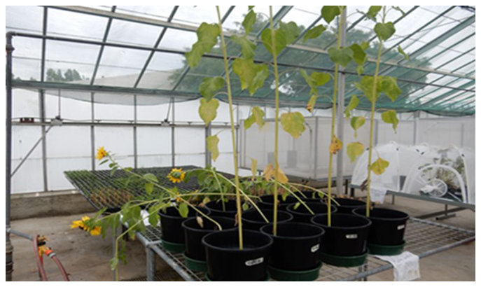
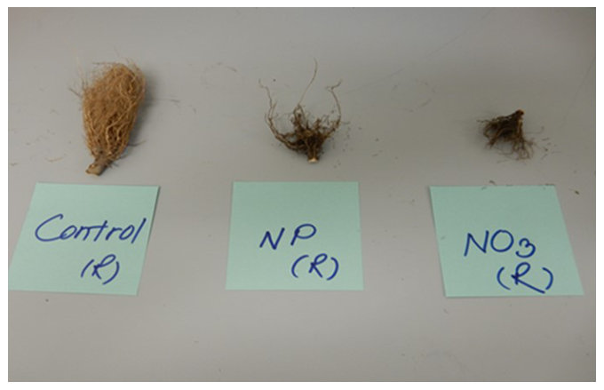
Sunflower plants exposed to AgNPs / Ag+ in a pot experiment with amended soil had fallen down by time of harvest (Figure 1) because of weak stems, probably caused by a deficiency of calcium (data shown in table 5). Silver from AgNPs or Ag+accumulated in root hair > root > leaf > stem > flower > seed (Table 2). The amount of Ag in sunflower leaves from exposure to Ag+in soil was 3-fold more than that accumulated on exposure to AgNPs and 6- fold more than the control. The seeds planted in soil amended with AgNPs showed a marked decrease in root size with only few roots having few root hairs (~50% less root hair), whereas the seeds grown in soil mixed with AgNO3 showed the smallest root size and ~75% less root hairs were evident compared to the control (Figure 2).
在添加处理土壤的盆栽试验中,暴露于AgNPs / Ag+的向日葵植株至收获时已倒伏(图1),原因是茎秆细弱,可能由钙缺乏所致(数据见表5)。来自AgNPs或Ag+的银累积顺序为根毛 > 根 > 叶 > 茎 > 花 > 种子(表2)。暴露于土壤Ag+的向日葵叶片中Ag含量是暴露于AgNPs时的3倍,是对照的6倍。播种于添加AgNPs土壤中的种子,其根系明显变小,仅有少量根具有少量根毛(根毛减少约50%);而生长于混有AgNO3土壤中的种子根系最小,与对照相比根毛减少约75%(图2)。
| Plant part植物部位 | Control对照 | AgNPsAgNPs | Ag~~+~~ |
|---|---|---|---|
| Root | 1.51 ± 0.45 | 17.83 ± 6.69 | 15.61 ± 1.96 |
| Root hairs | 2.65 ± 2 | 156 ± 9.69 | 182 ± 36.24 |
| Stem (S1) | 1.95 ± 1.09 | 3.75 ± 3.19 | 2.76 ± 1.16 |
| Stem (S2) | 1.32 ± 0.63 | 2.99 ± 2.67 | 1.3 ± 0.4 |
| Stem (S3) | 0.32 ± 0.04 | 0.76 ± 0.24 | 1.29 ± 0.6 |
| Stem (S4) | 0.26 ± 0.01 | 0.48 ± 0.54 | 1.27 ± 0.1 |
| Ag in whole stem | 3.85 ± 1.77 | 7.98 ±6.64 | 6.62 ± 2.26 |
| Leaf | 1.86 ± 0.24 | 2.75 ± 0.96 | 9.63 ± 4.94 |
| Flower | 0.31 ± 0.02 | 0.53 ± 0.14 | 1.09 ± 0.5 |
| Seed base | 0.42 ± 0.09 | 1.04 ± 0.59 | 0.85 ± 0.33 |
| Seeds | 0.32 ± 0.12 | 0.77 ± 0.21 | 1.04 ± 0.52 |
Exposure of sunflower plants to AgNPs/Ag+ in soil decreased the photosynthesis process resulting in generation of ROS, which damage chloroplast leading to growth retardation or death of the plants. In addition, a reduction in root size and lack of root hairs in the Ag-exposed plants was associated with a decrease in size of nearly all parts of the plant. This was more obvious in plants grown in soil amended with Ag+(as AgNO3) than AgNPs, which is consistent with the literature reports [32, 33]. Krizkova et al. [29] report a decrease of fresh weight in sunflower plants exposed to Ag+, and that this loss increased with time and exposure dose. These authors surmised that this weight loss was connected with an increase in metabolic activity due to very limited supply of the inorganic and organic compounds needed for plant growth. Krizkova et al. [29] reported that Ag changes water transport, which alters the anatomical structure of the root. The effects of Ag on sunflower roots they report include a decline in rhizodermal cells, which necrotized and were replaced by exodermal cells. Mazumdar [34] reports that when Ag particles enter root cells, they damage both the cell walls and vacuoles, which may contribute to increased entry of Ag into the plant including the root. Root damage could also be due to large Ag particles penetrating through small pores in the cell walls. Metal accumulation in plants is dependent on metal speciation, and Ag accumulates more in the root than other plant parts, followed by leaves and stem [33]. It is assumed that Ag will also accumulate in the seeds and if so, a food safety risk. Other factors that contribute to Ag accumulation in the root are solubility of Ag compounds, plant species and the culture medium [33].
向日葵植株暴露于土壤中的AgNPs/Ag+后,光合作用过程减弱,导致ROS生成,ROS损伤叶绿体,进而导致植株生长迟缓或死亡。此外,Ag暴露植株根系变小、缺乏根毛,这与植株几乎所有部位尺寸的减小相关。这一现象在添加Ag+(AgNO3形式)土壤中生长的植株中比在添加AgNPs土壤中更为明显,与文献报道一致[32, 33]。Krizkova et al. [29]报道,暴露于Ag+的向日葵植株鲜重下降,且这种损失随时间和暴露剂量的增加而加剧。这些作者推测,这种重量损失与代谢活动增强有关,原因在于植物生长所需的无机和有机化合物供应十分有限。Krizkova et al. [29]报道,Ag会改变水分运输,从而改变根的解剖结构。他们报道的Ag对向日葵根系的影响包括根表皮细胞减少,这些细胞坏死后被外皮层细胞取代。Mazumdar [34]报道,Ag颗粒进入根细胞后会损伤细胞壁和液泡,这可能促使更多Ag进入包括根在内的植物体。根系损伤也可能是由于较大的Ag颗粒穿透细胞壁上的小孔所致。植物中的金属累积取决于金属形态,Ag在根中的累积多于其他植物部位,其次为叶和茎[33]。推测Ag也会在种子中累积,若如此则构成食品安全风险。影响Ag在根中累积的其他因素包括Ag化合物的溶解度、植物种类和培养基质[33]。
The plants cultivated in soil amended with AgNPs or Ag+were not strong enough and collapsed (Figure 1) probably due to a deficiency of Ca as calcium pectate [35, 36]. When plants become deficient in Ca, the new tissues that are formed at root and shoot tips exhibit distorted growth due to improper cell formation. This may be the reason that the plants exposed to AgNPs/Ag+ curved at the upper parts of the plant resulting in tilting of the plant. The reduction of Ca concentration in the plants exposed to the two Ag forms (data shown on table 5) could also be attributed to a decrease in the expression of Ca channel proteins, which can lead to a reduction in Ca uptake. [37].
在添加AgNPs或Ag+的土壤中栽培的植株不够健壮而倒伏(图1),可能是由于以果胶酸钙形式存在的Ca缺乏所致[35, 36]。当植物缺Ca时,根尖和茎尖新形成的组织因细胞形成异常而表现出畸形生长。这可能是暴露于AgNPs/Ag+的植株上部弯曲、导致植株倾斜的原因。暴露于两种形态Ag的植株中Ca浓度降低(数据见表5),也可能归因于Ca通道蛋白表达下降,从而导致Ca吸收减少[37]。
3.2. Antioxidant enzymes, LPO, oxidase, peroxidases3.2. 抗氧化酶、LPO、氧化酶和过氧化物酶
Results are presented as the mean ± SE of four replicates and significance was set at P < 0.05.
结果以4次重复的均值 ± SE表示,显著性水平设为P < 0.05。
Compared to the control plants, leaves of sunflower plants grown in soil containing 150 mg/kg AgNPs or Ag+ (as AgNO3) respectively showed significantly increased catalase enzyme activity (> 8- and 12-fold), SOD activity (> 2- and 3-fold), and GPx enzyme activity (> 2- and 4-fold). GST enzyme activity in sunflower leaves exposed to Ag+ was significantly increased c. 2-fold compared to the control and plants exposed to AgNPs (Table 3).
与对照植株相比,生长于含150 mg/kg AgNPs或Ag+(AgNO3形式)土壤中的向日葵叶片,过氧化氢酶活性(分别 > 8倍和12倍)、SOD活性(分别 > 2倍和3倍)和GPx酶活性(分别 > 2倍和4倍)均显著升高。暴露于Ag+的向日葵叶片GST酶活性较对照及暴露于AgNPs的植株显著升高约2倍(表3)。
In sunflower leaves of plants exposed to AgNPs and Ag+ there were significant increases in MDA concentrations (0.3- and 0.7-fold more), ascorbate oxidase activity (2- and 4-fold more), pyrogallol peroxidase (6- and 11-fold more), and guaiacol peroxidase activ-
暴露于AgNPs和Ag+的植株向日葵叶片中,MDA浓度(分别增加0.3倍和0.7倍)、抗坏血酸氧化酶活性(分别增加2倍和4倍)、邻苯三酚过氧化物酶(分别增加6倍和11倍)以及愈创木酚过氧化物酶活
ity (~ 0.5 and 1.3-fold more) respectively compared to the control (Table 3).
性(分别增加约0.5倍和1.3倍)较对照均显著升高(表3)。
| Control对照 | AgNPsAgNPs | Ag~~+~~ | |
|---|---|---|---|
| Catalase | 0.24 ± 0.06~~a~~ | 4.24 ± 0.46~~b~~ | 6 ± 0.1~~c~~ |
| SOD | 10 ± 1.5~~a~~ | 21.11 ± 4~~b~~ | 32 ± 6~~c~~ |
| GST | 259.38 ± 0.29~~a~~ | 267 ± 23.08~~a~~ | 535 ± 147~~b~~ |
| GPx | 0.00032 ± 0.00005a | 0.0013 ± 0.0004b | 0.0022 ± 0.0002c |
| MDA conc. | 46 ± 1.63~~a~~ | 60 ± 1.24 b | 82 ± 1.12~~c~~ |
| Ascorbate oxidase | 8 ± 0.62~~a~~ | 15 ± 0.36~~b~~ | 18 ± 0.52~~c~~ |
| Pyrogallol peroxidase | 17 ± 2~~a~~ | 191 ± 31~~b~~ | 300 ± 56~~c~~ |
| Guaiacol peroxidase | 1.6 ± 0.41 a | 2.39 ± 0.11~~a~~ | 3.84 ± 0.26~~b~~ |
Means ± SE of four replicates. Means with different letters in a row are significantly different (P < 0.05).
数值为4次重复的均值 ± SE。同一行中标有不同字母的均值差异显著(P < 0.05)。
Reactive oxygen species are generated during the normal metabolism of eukaryotic cells [38] and may cause oxidative damage to macromolecules such as lipids, proteins, and DNA [39]. We analysed four antioxidant enzymes (SOD, CAT, GST, GPx) and LPO to evaluate the response to ROS in leaves of sunflowers exposed to 150 mg/kg AgNPs or Ag+in soil.
活性氧在真核细胞的正常代谢过程中产生[38],可能对脂质、蛋白质和DNA等大分子造成氧化损伤[39]。本研究分析了四种抗氧化酶(SOD、CAT、GST、GPx)和LPO,以评估暴露于土壤中150 mg/kg AgNPs或Ag+的向日葵叶片对ROS的响应。
In general, metal ions cause oxidative stress in sunflowers and generate ROS toxic species such as free radicals O•−2, OH• and non-radical like H2O2 and 1O2 that can cause degradation of plant proteins in addition to a reduction in number of chloroplasts in leaf tissue. Thus, ROS can affect a range of enzymes and activate the enzymatic antioxidant system, which responds to abiotic and biotic stressors [40, 41]. The antioxidant enzymes studied were CAT (catalyse dismutation of H2O2 to water and oxygen), SOD (frontline defence enzyme against ROS), GST (detoxification enzyme) and GPx (prevents damage from H2O2) [41].
一般而言,金属离子会引起向日葵氧化胁迫,并生成ROS毒性物质,如自由基O•−2、OH•以及非自由基H2O2和1O2,这些物质可导致植物蛋白降解,并使叶片组织中叶绿体数量减少。因此,ROS可影响多种酶并激活酶促抗氧化系统,该系统对非生物和生物胁迫因子作出响应[40, 41]。所研究的抗氧化酶包括CAT(催化H2O2歧化为水和氧气)、SOD(抵御ROS的第一道防线酶)、GST(解毒酶)和GPx(防止H2O2造成损伤)[41]。
Sunflower leaves exposed to the two Ag forms (AgNPs and Ag+) exhibited elevated CAT, SOD,
暴露于两种Ag形态(AgNPs和Ag+)的向日葵叶片表现出CAT、SOD、
GST and GPx activation, which is in agreement with [42], which found these enzymes were markedly elevated in sunflower leaf exposed to Ag+ more than AgNPs. Similar findings on increased CAT and GPx activities have been reported on exposure of Bacopa monnieri to 10 and 100 mg kg-1 Ag+, much higher than from exposure to the same concentration of AgNPs [11]. AgNPs sized 20 nm at a concentration of 10 mg L-1 significantly induced oxidative stress with an increase in ROS in a higher aquatic plant Spirodela polyrhiza. In S. polyrhiza exposed to 5 and 10 mg L-1 concentrations of 6-nm and 20-nm AgNPs particle sizes, SOD, CAT and guaiacol peroxidase were significantly increased [43], which also agrees with our results. Jiang et al. [43] reported that exposure of sunflowers to 6-nm AgNPs showed increased activity of these enzymes compared to 20-nm AgNPs particles, showing that the higher activity of the small sized particles was probably due to the relatively larger surface area.
GST和GPx活性升高,这与[42]的结果一致,该研究发现暴露于Ag+的向日葵叶片中这些酶的升高幅度明显大于暴露于AgNPs的叶片。已有类似报道表明,Bacopa monnieri暴露于10和100 mg kg-1 Ag+,后CAT和GPx活性升高,且远高于暴露于相同浓度AgNPs时的水平[11]。浓度为10 mg L-1、粒径20 nm的AgNPs在高等水生植物Spirodela polyrhiza中显著诱导了氧化胁迫并使ROS增加。在暴露于5和10 mg L-1浓度、粒径为6 nm和20 nm的AgNPs的S. polyrhiza中,SOD、CAT和愈创木酚过氧化物酶均显著升高[43],这也与本研究结果一致。Jiang et al. [43]报道,与20 nm AgNPs颗粒相比,暴露于6 nm AgNPs的向日葵中这些酶的活性更高,表明小粒径颗粒活性较高可能是由于其相对较大的比表面积。
Increased anthocyanins and chloroplast, which play a major role in photosynthesis, were increased in Arabidopsis thaliana exposed to AgNPs [44]. In addition, the plant tissues accumulated H2O2 and also exhibited an increase in nucleic acids and proteins, which could be detected by staining plant tissues with 3,3-diaminobenzidine. The plant leaf under stress exhibited a deep brown colour, compared with the control group. Excessive accumulation of ROS and subsequent LPO is identified by MDA measurement. Li et al. [44] reported that SOD activity in A. thaliana on exposure to AgNPs (0.1, 0.5, 1 mg L-1) significantly declined by 18.2% compared to the control, which is contrary to our findings. In contrast, CAT and POX in the AgNPs-treated group were induced by 2.1- and 1.1-fold respectively compared to the controls, which is in agreement with our findings. The reason for this could be the different plant types and AgNPs particle sizes. (30-nm in my study, and from 9 to 10 nm in [44]). Exposure of tobacco plants to 25-, 50-, 75-, 100- or 500-µM doses of AgNPs and Ag+ in milli Q water in an Erlenmeyer flask resulted in toxicity by Ag+> AgNPs [45], which is consistent with our observations in sunflowers grown in TSL soil, except for CAT activity which declined on exposure to AgNPs but increased on exposure to Ag+.
暴露于AgNPs的Arabidopsis thaliana中,在光合作用中起重要作用的花青素和叶绿体均有所增加[44]。此外,植物组织中累积了H2O2,核酸和蛋白质也有所增加,这可通过用3,3-二氨基联苯胺对植物组织染色检测到。与对照组相比,受胁迫的植物叶片呈深褐色。ROS的过量累积及随之发生的LPO可通过MDA测定加以识别。Li et al. [44]报道,A. thaliana暴露于AgNPs(0.1、0.5、1 mg L-1)后SOD活性较对照显著下降18.2%,这与本研究结果相反。相反,AgNPs处理组的CAT和POX分别较对照被诱导升高2.1倍和1.1倍,这与本研究结果一致。其原因可能在于植物种类和AgNPs粒径不同(本研究为30 nm,[44]中为9~10 nm)。将烟草植株置于锥形瓶中,以milli Q水中25、50、75、100或500 µM剂量的AgNPs和Ag+处理,其毒性表现为Ag+> AgNPs[45],这与本研究在TSL土壤中生长的向日葵的观察结果一致,但CAT活性例外:暴露于AgNPs时CAT活性下降,而暴露于Ag+时升高。
Tomato plants exposed to 0, 25, 50, 75, and 100 mg L-1 of 20-nm AgNPs resulted in a linear doseresponse increase in all amino acids and also the activity of antioxidant enzymes CAT, SOD, POX in shoot and root at 50 mg/L Ag but total soluble protein decreased [46], all of which agrees with our results. However, at 100 mg L-1 AgNPs, SOD activity declined probably due to unspecified enzyme degradation [47] or due to binding of Ag to the active centre of the SOD enzyme [48], the activity of which was higher in the shoot than roots. The SOD and CAT activity increased in castor oil seedlings exposed to AgNPs/Ag+ [49], which is in agreement with our data. Heavy metals such as Cu and Zn also induced CAT, SOD, POX in castor beans Ricinus communis [49].
番茄植株暴露于0、25、50、75和100 mg L-1的20 nm AgNPs后,所有氨基酸均呈线性剂量-效应增加;在50 mg/L Ag时,地上部和根中抗氧化酶CAT、SOD、POX的活性也升高,但总可溶性蛋白下降[46],这些均与本研究结果一致。然而,在100 mg L-1 AgNPs时,SOD活性下降,可能是由于非特异性的酶降解[47],或是由于Ag与SOD酶活性中心结合[48];SOD活性在地上部高于根部。暴露于AgNPs/Ag+的蓖麻幼苗中SOD和CAT活性升高[49],与本研究数据一致。Cu和Zn等重金属也能诱导蓖麻Ricinus communis中的CAT、SOD和POX[49]。
The effect of AgNPs on sunflower leaf GST activity was minimal but Ag+ increased GST compared to the control. GST is a detoxification enzyme involved in the metabolism of many chemicals including pesticides and heavy metals. It catalyses the conjugation of electrophilic molecules with reduced glutathione (GSH) to more water soluble and less toxic metabolites [50]. The GSH conjugation is the first step of the mercapturic acid pathway, which is an important detoxification process [51]. GST is induced and able to detoxify chlorophenol in plants grown in polluted soil [52] including on exposure to high concentrations of Cd [53].
AgNPs对向日葵叶片GST活性的影响甚微,但与对照相比,Ag+使GST升高。GST是一种解毒酶,参与包括农药和重金属在内的多种化学物质的代谢。它催化亲电分子与还原型谷胱甘肽(GSH)结合,生成水溶性更强、毒性更低的代谢产物[50]。GSH结合是巯基尿酸途径的第一步,该途径是一个重要的解毒过程[51]。在污染土壤中生长的植物中,GST被诱导并能够对氯酚进行解毒[52],暴露于高浓度Cd时亦是如此[53]。
A significant increase in GPx was observed in sunflower leaf in plants exposed to AgNPs and Ag+ compared to the controls. The GPx activity in plants depends on physiological and genetic status, time interval, type and concentration of exposure to pollutants including heavy metals and is a response to oxidative stress [54]. GPx catalyses the reduction of H2O2 via GSH to protect plant cells from oxidative stress [38] not only caused by heavy metals but also by salinity and drought conditions [55].
与对照相比,暴露于AgNPs和Ag+的植株向日葵叶片中GPx显著升高。植物中的GPx活性取决于生理和遗传状态、时间间隔以及包括重金属在内的污染物的暴露类型和浓度,是对氧化胁迫的一种响应[54]。GPx通过GSH催化H2O2的还原,保护植物细胞免受氧化胁迫[38],这种氧化胁迫不仅由重金属引起,也可由盐渍和干旱条件引起[55]。
Nickle (Ni) is a heavy metal that cause oxidative stress in Amaranthus paniculatus with a resultant increase in LPO, GPx, SOD and CAT [56]. However, the authors reported that antioxidant enzymes including GPx in roots was progressively inhibited by increased Ni concentration in the growth solution but that a marked increase in GPx and SOD activity occurred in leaves. There are several publications [12, 57-60] on oxidative stress in plants with resultant increase in ROS, LPO and GPx. The exposure of sunflowers to AgNPs and Ag+ resulted in an increase not only in GPx but also other POX such as pyrogallol peroxidase and guaiacol peroxidase.
镍(Ni)是一种可引起Amaranthus paniculatus氧化胁迫的重金属,导致LPO、GPx、SOD和CAT升高[56]。然而,作者报道,随着生长溶液中Ni浓度的增加,根中包括GPx在内的抗氧化酶受到逐渐增强的抑制,而叶片中GPx和SOD活性则显著升高。已有多篇文献[12, 57-60]报道了植物氧化胁迫及由此导致的ROS、LPO和GPx升高。向日葵暴露于AgNPs和Ag+后,不仅GPx升高,邻苯三酚过氧化物酶和愈创木酚过氧化物酶等其他POX也升高。
Exposure of plants to external stress induces LPO. Sunflower plants exposed to AgNPs and Ag+ resulted in a significant increase in MDA in the leaf with the effects due to Ag+ more than AgNPs. In A. thaliana exposed to AgNPs and Ag+, tissues accumulated ROS and resulted in LPO in the leaf, which was quantified by MDA [44]. A similar effect was observed in tomato plants, with a concentrationdependent increase in MDA on exposure to Ag [61]. Exposure of the aquatic plant Spirodela polyrhiza to 5 mg L-1 AgNPs in 10% Hoagland solution resulted in a significant increase in LPO compared to the control [62]. MDA was also significantly increased in leaves of the tobacco plant exposed to 500 µM AgNPs and to 100 µM Ag+[45]. Thus, the changes in MDA observed in the leaves when the tobacco plant is exposed to AgNPs/Ag+ are similar to the ones observed in our study with sunflower leaves. The main effects of heavy metal ions on plants include an increase in LPO and lipoxygenase activity, and an associated decrease in chlorophyll and GSH [7]. Ascorbate oxidase is also an antioxidant defence enzyme, which can resist ROS. It plays a major role in detoxifying H2O2 in plant cells. The ascorbate oxidase catalyses the conversion of H2O2 to water by acting as a specific electron donor [63]. An increase in the POX enzyme on exposure to AgNPs and Ag+ has been reported [64]. The POX used in our study were pyrogallol peroxidase and guaiacol peroxidase
植物暴露于外界胁迫会诱发LPO。暴露于AgNPs和Ag+的向日葵植株叶片中MDA显著增加,且Ag+的效应大于AgNPs。在暴露于AgNPs和Ag+的A. thaliana中,组织累积ROS并导致叶片发生LPO,其程度以MDA定量[44]。在番茄植株中也观察到类似效应,暴露于Ag后MDA呈浓度依赖性增加[61]。水生植物Spirodela polyrhiza暴露于10% Hoagland溶液中5 mg L-1的AgNPs后,LPO较对照显著增加[62]。暴露于500 µM AgNPs和100 µM Ag+的烟草植株叶片中MDA也显著增加[45]。因此,烟草植株暴露于AgNPs/Ag+时叶片中MDA的变化与本研究在向日葵叶片中观察到的变化相似。重金属离子对植物的主要影响包括LPO和脂氧合酶活性升高,以及相应的叶绿素和GSH下降[7]。抗坏血酸氧化酶也是一种能够抵御ROS的抗氧化防御酶,在植物细胞中H2O2的解毒过程中起重要作用。抗坏血酸氧化酶作为特异性电子供体,催化H2O2转化为水[63]。已有报道表明,暴露于AgNPs和Ag+后POX酶升高[64]。本研究所用的POX为邻苯三酚过氧化物酶和愈创木酚过氧化物酶。
Exposure of Bacopa monnieri to AgNPs produced more pyrogallol peroxidase than in plants exposed to Ag+ [11] along with an increase in the TPC and a decrease in protein and carbohydrate. In our study, the increase in pyrogallol peroxidase on exposure of the sunflower plant to Ag+ was significantly higher than by AgNPs, which is similar to the finding in the tobacco plants exposed to 50, 75, 100 and 500 µM [45] but different to that reported by [11] in B. monnieri. Guaiacol peroxidase activity on exposure of sunflower to the two Ag forms showed significant increase in the plants exposed to Ag+ only and not AgNPs compared to the control, which is in agreement with the observations of [61] who exposed the tomato plant to AgNPs (6 nm particle size) and Ag+ and observed a significant increase in guaiacol peroxidase activity and also the findings in the aquatic plant Spirodela polyrhiza exposed to AgNPs [62].
Bacopa monnieri暴露于AgNPs时产生的邻苯三酚过氧化物酶多于暴露于Ag+的植株[11],同时TPC增加,蛋白质和碳水化合物减少。本研究中,向日葵暴露于Ag+时邻苯三酚过氧化物酶的增加显著高于暴露于AgNPs时,这与暴露于50、75、100和500 µM的烟草植株中的发现相似[45],但与[11]在B. monnieri中的报道不同。向日葵暴露于两种Ag形态后,与对照相比,仅暴露于Ag+的植株愈创木酚过氧化物酶活性显著升高,而暴露于AgNPs的植株则未显著升高;这与[61]的观察结果一致,该研究将番茄植株暴露于AgNPs(粒径6 nm)和Ag+,观察到愈创木酚过氧化物酶活性显著升高;也与暴露于AgNPs的水生植物Spirodela polyrhiza中的发现一致[62]。
3.3. Total phenolic compounds and Total soluble carbohydrates3.3. 总酚类化合物与总可溶性碳水化合物
The TPC concentration was significantly increased in the leaves of sunflower plants exposed to Ag+ > AgNPs > control. A similar observation has been recorded on exposure of B. monnieri to heavy metals [11] and also that phenolic derivatives can act as a metal stress chelator, as an antioxidant to reduce ROS and LPO. These authors reported a reduction in protein and carbohydrate concentrations also [11]. Total phenolic compounds increased by 0.3- and 1- fold, whereas total soluble carbohydrates decreased by 20% and 35%, respectively compared to the control.
向日葵叶片中TPC浓度显著升高,顺序为Ag+ > AgNPs > 对照。在B. monnieri暴露于重金属时也观察到类似现象[11],且酚类衍生物可作为金属胁迫螯合剂,并作为抗氧化剂降低ROS和LPO。这些作者还报道了蛋白质和碳水化合物浓度的降低[11]。与对照相比,总酚类化合物分别增加了0.3倍和1倍,而总可溶性碳水化合物分别降低了20%和35%。
Total soluble carbohydrate concentration in sunflower leaves significantly declined following exposure of plants to 150 mg kg-1 of AgNPs and Ag+ in soil for 53 days and this was most marked in the plants exposed to Ag+. Similar results have been reported in B. monnieri exposed to 10, 100 ug/kg of AgNPs and 10, 100 mg kg-1 of Ag+ with a slow decline during the first 10 days of cultivation and a sharp decrease in TSC thereafter [11], which agreed with our findings. Similarly, Salama [10] reported that the exposure of corn (Zea mays) and common bean (Phaseolus vulgaris) to doses up 60 mg kg-1of AgNPs caused an increase in TSC, but at higher AgNPs concentrations, such as 80 mg kg-1, the TSC concentration decreased significantly. Application of colloidal AgNPs (1 mg/ ml) as a foliar spray dose of 50 ml/L increased TSC and also improved the appearance of sunflower leaves [65]
植株暴露于土壤中150 mg kg-1的AgNPs和Ag+ 53天后,向日葵叶片中总可溶性碳水化合物浓度显著下降,且在暴露于Ag+的植株中最为明显。在暴露于10、100 ug/kg AgNPs和10、100 mg kg-1 Ag+的B. monnieri中也报道了类似结果:栽培最初10天内TSC缓慢下降,此后急剧下降[11],这与本研究结果一致。同样,Salama [10]报道,玉米(Zea mays)和菜豆(Phaseolus vulgaris)暴露于最高60 mg kg-1的AgNPs时TSC增加,但在较高AgNPs浓度(如80 mg kg-1)下TSC浓度显著下降。以50 ml/L的剂量叶面喷施胶体AgNPs(1 mg/ ml)可提高TSC,并改善向日葵叶片的外观[65]。
3.4. Vitamins A, E and C3.4. 维生素A、E和C
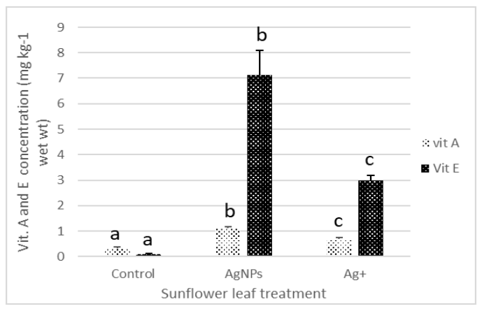
Vitamin A concentration in the leaves of sunflower plants exposed to AgNPs and Ag+were increased by 1- and 0.3-fold, and vitamin E increased by 6.8- and 2.8-fold, respectively compared to the control (Figure 4). Vitamin C concentrations in leaves exposed to AgNPs and Ag+increased by 25% (P < 0.05) and 21% respectively compared to the control.
与对照相比,暴露于AgNPs和Ag+的向日葵叶片中维生素A浓度分别增加了1倍和0.3倍,维生素E分别增加了6.8倍和2.8倍(图4)。暴露于AgNPs和Ag+的叶片中维生素C浓度较对照分别增加了25%(P < 0.05)和21%。
Exposure of sunflower plant to 150 mg kg-1 AgNPs or Ag+resulted ina significant increase in vitamin A and E concentrations, with AgNPs > Ag+> control. The exposure of sunflower plants to AgNPs and Ag+ stimulates the production of ascorbate oxidase due to ROS production, which results in the production of L -ascorbic acid. Vitamins A and E (alpha-tocopherol) act as a defence in addition to ascorbate oxidase, which oxidises L-ascorbic acid to dehydroascorbic acid, and therefore the amount of dehydroascorbic acid is increased, and the total ascorbic acid is decreased [66]. A significant increase in the antioxidant vitamins A and E in the bean plant (Phaseolus vulgarus) occurred following exposure to heavy metals Pb (1.5, 2, 2.5 mM), Cu (0.1, 0.2, 0.3 mM), Cd (0.05, 0.06 and 0.08 mM) and Hg (0.02, 0.04 and 0.06) for 10 days [67], which is in agreement with our findings.
向日葵植株暴露于150 mg kg-1 AgNPs或Ag+后,维生素A和E浓度显著增加,顺序为AgNPs > Ag+> 对照。向日葵植株暴露于AgNPs和Ag+后,由于ROS的产生而刺激抗坏血酸氧化酶的生成,进而生成L-抗坏血酸。维生素A和E(α-生育酚)与抗坏血酸氧化酶共同发挥防御作用,抗坏血酸氧化酶将L-抗坏血酸氧化为脱氢抗坏血酸,因此脱氢抗坏血酸含量增加,而总抗坏血酸含量降低[66]。菜豆(Phaseolus vulgarus)暴露于重金属Pb(1.5、2、2.5 mM)、Cu(0.1、0.2、0.3 mM)、Cd(0.05、0.06和0.08 mM)和Hg(0.02、0.04和0.06)10天后,抗氧化维生素A和E显著增加[67],这与本研究结果一致。
3.5. Chlorophyll and total carotenoids3.5. 叶绿素与总类胡萝卜素
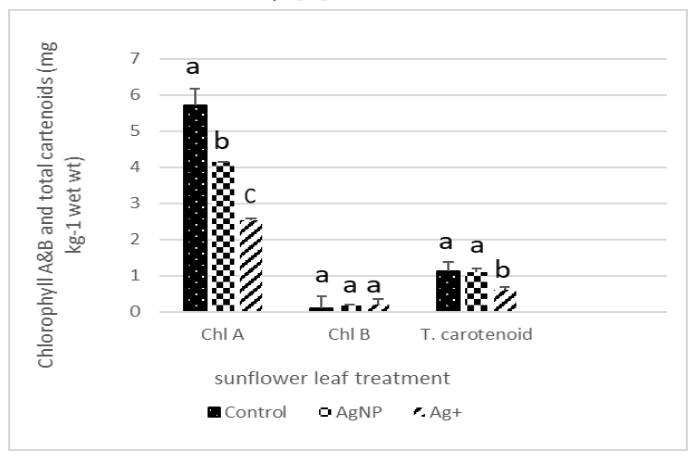
Sunflower leaf chlorophyll A in plants grown in soil containing 150 mg kg-1 Ag as AgNPs and Ag+ (as AgNO3) decreased by 30% and 40% respectively. Chlorophyll B was not significantly affected. Total carotenoid concentration in the sunflower leaves exposed to Ag+ decreased by ~ 50% compared to the control (Figure 3).
生长于含150 mg kg-1 Ag(以AgNPs和Ag+(AgNO3形式))土壤中的植株,其向日葵叶片叶绿素A分别下降了30%和40%。叶绿素B未受到显著影响。暴露于Ag+的向日葵叶片中总类胡萝卜素浓度较对照下降约50%(图3)。
Leaf pigments include chlorophyll A and B and total carotenoids, which are essential for photosynthesis and hence plant growth [68]. Silver nanoparticles are taken up by plants into intracellular spaces and transported inside the plant through the plasmodesmata of root cells, then pass through shoots and accumulate in leaves causing inhibition of chlorophyll. The ratio between chlorophyll A and chlorophyll B indicates the response to light and shade conditions and is used as a sensitive biomarker of pollution and environmental stress [69]. Carotenoids are located in chloroplast and with chlorophyll are intricately involved in the photosynthetic process. In addition, they can protect chlorophyll and the thylakoid membrane (a sheet like membrane-bound structure in chloroplast that plays a role in light-dependent photosynthesis) from peroxidative damage [25]. Carotenoids dissolve in non-polar and polar solvents such as diethyl ether and methanol. Hence, we selected methanol for extraction of chlorophyll and total carotenoids in a one-step extraction with good recovery; besides methanol is easier to handle. Mazumdar [34] reported that using 1000 µg/ ml AgNPs in Hoagland’s nutrient solution caused a significant inhibition in both chlorophyll types and total carotenoids in Vigna radiata and Bactris campestris, which is in agreement with our findings There are several reports that AgNPs inhibit plant growth by inhibition of chlorophyll formation and total soluble protein [70, 71] . However, Qian [12] showed that AgNPs inhibited the growth of A. thaliana by disruption of the thylakoid membrane structure and thereby decreased the chlorophyll concentration. In our study, we did not investigate the impact of AgNPs on thylakoid membrane structure, but we agree that chlorophyll concentration is reduced on exposure to AgNPs. In contrast, Qian et al. [12] suggested that Ag+ as AgNPs did not profoundly affect chloroplast structure but could alter the transcription of antioxidants and aquaporin genes, indicating that AgNPs can change the balance between oxidant and antioxidant activity thereby affecting the homoeostasis of the A. thaliana plant. The decline in chlorophyll content in plants exposed to heavy metals has been reported in several studies, including a decrease in the ratio of chlorophyll A to chlorophyll B on exposure to Cu and Hg stress with chlorophyll B responding more to Cu, while a decrease in this ratio was observed in plants exposed to other heavy metals such as Cd and Pb [54].
叶片色素包括叶绿素A、叶绿素B和总类胡萝卜素,它们是光合作用进而是植物生长所必需的[68]。银纳米颗粒被植物吸收进入细胞间隙,并经根细胞的胞间连丝在植物体内转运,随后通过地上部并在叶片中积累,从而抑制叶绿素。叶绿素A与叶绿素B的比值反映植物对光照和遮荫条件的响应,可用作污染和环境胁迫的敏感生物标志物[69]。类胡萝卜素位于叶绿体中,与叶绿素共同密切参与光合作用过程。此外,类胡萝卜素还可保护叶绿素和类囊体膜(叶绿体中一种片层状膜结构,在光依赖性光合作用中发挥作用)免受过氧化损伤[25]。类胡萝卜素可溶于乙醚和甲醇等非极性和极性溶剂。因此,本研究选用甲醇一步提取叶绿素和总类胡萝卜素,回收率良好;此外,甲醇也更易于操作。Mazumdar [34]报道,在Hoagland营养液中施用1000 µg/ ml AgNPs显著抑制了Vigna radiata和Bactris campestris中两种叶绿素及总类胡萝卜素的含量,这与本研究结果一致。已有多项报道表明,AgNPs通过抑制叶绿素形成和总可溶性蛋白合成而抑制植物生长[70, 71]。然而,Qian [12]发现,AgNPs通过破坏类囊体膜结构抑制A. thaliana的生长,从而降低叶绿素浓度。本研究未考察AgNPs对类囊体膜结构的影响,但认同暴露于AgNPs会导致叶绿素浓度降低。与此相反,Qian et al. [12]认为,以AgNPs形式存在的Ag+对叶绿体结构影响不大,但可改变抗氧化物质和水通道蛋白基因的转录,表明AgNPs可改变氧化与抗氧化活性之间的平衡,进而影响A. thaliana植株的稳态。多项研究报道了暴露于重金属的植物叶绿素含量下降,包括在Cu和Hg胁迫下叶绿素A与叶绿素B的比值降低,且叶绿素B对Cu的响应更强;而在暴露于Cd和Pb等其他重金属的植物中也观察到该比值的下降[54]。
Carotenoids are a partner pigment in photosynthesis. Carotenoids also act as an antioxidant non-enzymatic pigment that defends chlorophyll and cell membranes against ROS produced on exposure to different heavy metals and environmental stress [72-74]. A role of this pigment is to reduce chlorophyll concentration to protect against chloroplast damage [74]. Decline in carotenoid content indicates heavy metal toxicity, while its enhancement reflects detoxification of ROS species [75, 76]. Our results that chlorophyll and total carotenoids are inhibited in sunflower leaves on exposure of the sunflower plant to AgNPs and Ag+in soil are confirmed by [7].
类胡萝卜素是光合作用中的辅助色素。类胡萝卜素还作为一种非酶抗氧化色素,保护叶绿素和细胞膜免受暴露于各种重金属和环境胁迫时产生的ROS的伤害[72-74]。该色素的作用之一是降低叶绿素浓度,以防止叶绿体受损[74]。类胡萝卜素含量下降表明重金属毒性,而其含量升高则反映了对ROS的解毒作用[75, 76]。本研究发现,向日葵植株暴露于土壤中的AgNPs和Ag+后,其叶片中叶绿素和总类胡萝卜素受到抑制,这一结果得到了文献[7]的证实。
| Control对照 | AgNPsAgNPs | Ag+ | |
|---|---|---|---|
| Total protein (mg/ kg wet wt). | 20.18 ± 0.77~~a~~ | 16.74 ± 0.95~~b~~ | 14.14 ± 0.97~~c~~ |
| Total phenolic compounds (mg equivalent to gallic acid wet wt). | 0.151± 0.01~~a~~ | 0.203 ± 0.002 b | 0.294 ± 0.02~~c~~ |
| Total soluble carbohydrates (g/ kg wet wt). | 168 ± 9.65~~a~~ | 142 ± 8.8~~b~~ | 109 ± 12.6 |
| Urease (mmol/ g wet wt) | 3.03 ± 0.3~~a~~ | 4.27 ± 0.79~~b~~ | 6 ± 1.21~~c~~ |
| Vitamin C (mg/kg wet wt) | 117.16 ± 9~~a~~ | 166.46 ± 16.8~~b~~ | 147. 91 ± 2.77~~c~~ |
Means ± SE of four replicates. Means with different letters in a row are significantly different (P < 0.05).
数值为4次重复的均值 ± SE。同一行中标有不同字母的均值差异显著(P < 0.05)。
3.6. Urease concentration3.6. 脲酶浓度
Urease levels increased significantly with the Ag+ treatment (1-fold more) but were not significantly different from the control with AgNPs treatment (~0.4 fold more) (Table 4).
Ag+处理使脲酶水平显著升高(增加1倍),而AgNPs处理与对照相比差异不显著(增加约0.4倍)(表4)。
Urease is a crucial plant enzyme that catalyses the hydrolysis of urea to form ammonia and carbon dioxide [77]. In our study, there was a significant increase in urease activity in sunflower leaves with the increase being most marked in plants exposed to Ag+ more than AgNPs more than control.
脲酶是一种重要的植物酶,可催化尿素水解生成氨和二氧化碳[77]。本研究中,向日葵叶片脲酶活性显著升高,其升高幅度依次为暴露于Ag+的植株 > 暴露于AgNPs的植株 > 对照。
3.7. Total protein3.7. 总蛋白
The total protein concentrations in sunflower leaves exposed to AgNPs and Ag+(as AgNO3) were ~ 12.5% and 25% lower respectively compared to the control. Proteins are large biomolecules consisting of a series of amino acids. In plants, protein has five main functions: plant growth, role in hormones and enzymes, immune function, and in energy production. Exposure of sunflower plants to AgNPs and Ag+ resulted in a reduction of protein in sunflower leaf with Ag+> AgNPs> control. Exposure of sunflower plants to 0 (control), 0.1, 0.5- and 1-mM Ag+ cultivated in an environmental test chamber (MLR-350 H, Sanyo, Japan) showed that protein content in the shoot > root indicative of transport of proteins from roots to shoots [29]. A reduced protein concentration in both the shoot more than root exposed to heavy metals has been reported [41]. A similar decline of up to 70% protein has been observed in the tomato plant leaf exposed to 50 µM Cd in soil compared to the controls and this was accompanied by an accumulation of ammonium ions [61]. In the aquatic plant Spirodela polyrhiza, the effect of 6-nm AgNPs of 5 mg/ L increased total soluble protein content [43]. The increase in total soluble protein was much higher when the plant was exposed to 10 mg L-1 20-nm sized AgNPs [43]. This is in contrast to our finding of a decline in sunflower leaf protein in plants exposed to 150 mg/kg Ag and this was most marked in the plants exposed to Ag+more than AgNPs. This could be due to the smaller particles size and the lower Ag dose used by [43]. Heavy metals such as Pb, Cd, Mn, Co, Cr, and Co also cause significant inhibition of total soluble protein and plant growth in Zea mays [78], which also agreed with our results. On exposure of Bacopa monnieri to AgNPs, the leaf protein concentration declined over time, which agreed with our study [11]. These authors also added that the decrease in protein concentration in B. monnieri was accompanied by an increase in TPC on exposure to heavy metals. The reduction in total amount of soluble protein when exposed to heavy metals could be due to an increase in protease activity [79] or because of the diverse structural alteration and thereby denaturing the protein [80].
与对照相比,暴露于AgNPs和Ag+(AgNO3)的向日葵叶片总蛋白浓度分别降低约12.5%和25%。蛋白质是由一系列氨基酸组成的大分子生物分子。在植物中,蛋白质具有五项主要功能:促进植物生长、参与激素和酶的作用、免疫功能以及能量生产。向日葵植株暴露于AgNPs和Ag+后,叶片蛋白质含量降低,降幅依次为Ag+> AgNPs> 对照。在环境试验箱(MLR-350 H, Sanyo, Japan)中培养并暴露于0(对照)、0.1、0.5和1 mM Ag+的向日葵植株,其地上部蛋白质含量 > 根部,表明蛋白质由根部向地上部转运[29]。已有报道指出,暴露于重金属后地上部和根部的蛋白质浓度均降低,且地上部降幅大于根部[41]。在土壤中暴露于50 µM Cd的番茄叶片中也观察到类似现象,与对照相比蛋白质下降达70%,并伴随铵离子的积累[61]。在水生植物Spirodela polyrhiza中,5 mg/ L的6 nm AgNPs使总可溶性蛋白含量增加[43]。当该植物暴露于10 mg L-1的20 nm AgNPs时,总可溶性蛋白增幅更大[43]。这与本研究中暴露于150 mg/kg Ag的向日葵叶片蛋白质下降的结果相反,且该下降在暴露于Ag+的植株中比暴露于AgNPs的植株更为明显。这可能是由于文献[43]所用颗粒粒径更小、Ag剂量更低所致。Pb、Cd、Mn、Co、Cr和Co等重金属也会显著抑制Zea mays的总可溶性蛋白和植物生长[78],这同样与本研究结果一致。Bacopa monnieri暴露于AgNPs后,其叶片蛋白质浓度随时间下降,与本研究一致[11]。这些作者还指出,B. monnieri暴露于重金属时,蛋白质浓度的降低伴随着TPC的增加。暴露于重金属时可溶性蛋白总量的减少,可能是由于蛋白酶活性升高[79],或由于蛋白质发生多种结构改变进而变性[80]。
4CONCLUSIONS结论
Exposure of sunflower plants to AgNPs and Ag+(as AgNO3) caused oxidative stress similar to that caused by other heavy metals such as Cd, Pb, Hg, and Ni. The stress of exposure to such heavy metals can lead to ROS, LPO and elevated defence enzymes (CAT, SOD, GST, and GPx) activities in addition to increased activity of POX (pyrogallol peroxidase and guaiacol peroxidase), and antioxidant vitamins (Vitamin A, E, and C) and TPC. The increase in TPC was accompanied by a decrease in total soluble protein. Silver accumulated in the sunflower plant with the concentration in the root more than leaf more than stem. Silver nanoparticles can alter the transcription of antioxidants and oxidants, causing an imbalance in homoeostasis of the plant. The major impacts of AgNPs and Ag+ on the sunflower plant are the reduction of total protein, inhibition of chlorophyll synthesis through damage to chloroplast and TSC in the leaf, reduction in root size, loss of root hair and inhibition of stem length, all of which together can lead to a reduction in growth and yield of the crops. The ratio of chlorophyll A to chlorophyll B could be used as a sensitive biomarker of exposure to pollution and environmental stress. Overall, the responses of the plant parameters measured in this study were most marked in the sunflower plants exposed to Ag+ > AgNPs.
向日葵植株暴露于AgNPs和Ag+(AgNO3)后产生的氧化胁迫,与Cd、Pb、Hg和Ni等其他重金属引起的氧化胁迫相似。暴露于此类重金属的胁迫可导致ROS、LPO及防御酶(CAT、SOD、GST和GPx)活性升高,同时POX(邻苯三酚过氧化物酶和愈创木酚过氧化物酶)活性、抗氧化维生素(维生素A、E和C)及TPC也随之增加。TPC的增加伴随着总可溶性蛋白的减少。银在向日葵植株中积累,其浓度依次为根 > 叶 > 茎。银纳米颗粒可改变抗氧化物质和氧化物质的转录,导致植物稳态失衡。AgNPs和Ag+对向日葵植株的主要影响包括:总蛋白减少,因叶绿体受损而抑制叶片叶绿素合成和TSC,根系变小,根毛脱落以及茎长受抑制;上述影响共同作用可导致作物生长和产量下降。叶绿素A与叶绿素B的比值可用作污染和环境胁迫暴露的敏感生物标志物。总体而言,本研究所测植物参数的响应强度依次为暴露于Ag+ > AgNPs的向日葵植株。
| Element元素 | Ca | AgNO3) caused oxidative stress similar to that causedAgNO3)引起的氧化胁迫,与……引起的相似 |
|---|---|---|
| Part 1: Root | by other heavy metals such as Cd, Pb, Hg, and Ni. The stress of exposure to such heavy metals can lead | |
| Control | 5129 ± 422~~a~~ | to ROS, LPO and elevated defence enzymes (CAT, |
| AgNPs | 4077 ± 273~~ab~~ | SOD, GST, and GPx) activities in addition to in- |
| Ag~~+~~ | 3135± 110~~b~~ | creased activity of POX (pyrogallol peroxidase and |
| Part 2: Stem (S-1) | guaiacol peroxidase), and antioxidant vitamins (Vitamin A E and C) and TPC. The increase in TPC | |
| Control | 6974 ± 568~~a~~ | , , was accompanied by a decrease in total soluble pro- |
| AgNPs | 5789 ± 174~~a~~ | tein. Silver accumulated in the sunflower plant with |
| Ag~~+~~ | 5555 ± 301~~a~~ | the concentration in the root more than leaf more |
| Part 3: Stem (S-2) | than stem. Silver nanoparticles can alter the transcrip- | |
| Control | 10684 ± 177~~a~~ | tion of antioxidants and oxidants, causing an imbal- ance in homoeostasis of the plant. The major impacts |
| AgNPs | 57761 ± 291~~b~~ | of AgNPs and Ag+on the sunflower plant are the re- |
| Ag~~+~~ | 7502 ± 188~~c~~ | duction of total protein, inhibition of chlorophyll syn- |
| Part 3: Stem (S-3) | thesis through damage to chloroplast and TSC in the | |
| Control | 9369 ± 716~~a~~ | leaf, reduction in root size, loss of root hair and inhi- bition of stem length, all of which together can lead |
| AgNPs | 7777 ± 374~~ab~~ | to a reduction in growth and yield of the crops. The |
| Ag~~+~~ | 6041 ± 365~~b~~ | ratio of chlorophyll A to chlorophyll B could be used |
| Part 3: Stem (S-4) | as a sensitive biomarker of exposure to pollution and | |
| Control | 10860 ± 117~~a~~ | environmental stress. Overall, the responses of the plant parameters measured in this study were most |
| AgNPs | 9953 ± 722~~b~~ | marked in the sunflower plants exposed to Ag+> |
| Ag~~+~~ | 7260 ± 330~~c~~ | AgNPs. |
| Part 4: Leaves | ||
| Control | 40939 ± 830~~a~~ | The security risk of sunflower seeds after exposure to AgNPs and Ag+ increased and authorities must con- |
| AgNPs | 28815 ± 2001~~b~~ | trol the discharge of the waste of the factory used |
| Ag~~+~~ | 27044 ± 1692~~b~~ | AgNPs in their products and the waste of municipali- |
| Part 5: Flower | ty which contains AgNPs before use this waste as | |
| Control | 11080 ± 830~~a~~ | fertilizer. |
| AgNPs | 8903 ± 117~~b~~ | 5. ACKNOWLEDGMENTS |
| Ag~~+~~ | 4815 ± 262~~c~~ | This work was supported by Lincoln University and |
| Part 6: Seed | Landcare Research, Lincoln, New Zealand. The tech- | |
| Control | 15926 ± 1003~~a~~ | nical assistance of Mr. Martin Webley is gratefully kldd |
| AgNPs | 10274 ± 969~~ab~~ | acnowege. |
| Ag~~+~~ | 53041 ± 1256~~b~~ | 6 CONFLICT OF INTEREST |
6CONFLICT OF INTEREST利益冲突
The authors declare that they have no conflicts of interest.
作者声明不存在利益冲突。
REFERENCES参考文献
[1] Dakal, T.C., et al., Mechanistic basis of antimicrobial actions of silver nanoparticles. Frontiers in Microbiology. 2016.7:1831.
[2] Graf, C., et al., A general method to coat colloidal padticles with silica. Langmuir, 2003;19(17):6693– 7003. View Article
[3] Gatto, M A., Nassem, S., Arafat, M. Y., Mahmoud, D. A., Qasim, k., Zubai, S. Physiochemical properties of nanomaterials implications in associated toxic manifestation. BioMed Research, 2014;2: 1 – 8. [4] Anjum, N.A., et al., Silver nanoparticles in soil– plant systems. Journal of Nanoparticle Research. 2013.15(9):1896.
[5] Sairam, R., D. Singh, and G. Srivastava, Changes in activities of antioxidant enzymes in sunflower leaves of different ages. Biologia Plantarum. 2003.47(1):61.
[6] Stoikou, V., et al., Μetal uptake by sunflower (Helianthus annuus) irrigated with water polluted with chromium and nickel. Foods. 2017;6(7):51. PMid:28714908 View Article PubMed/NCBI
[7] Gallego, S.M., M.P. Benavídes, and M.L. Tomaro, Effect of heavy metal ion excess on sunflower leaves: evidence for involvement of oxidative stress. Plant Science, 1996. 121(2):151–9. 04528-1 View Article
[8] Leela, A. and M. Vivekanandan, Tapping the unexploited plant resources for the synthesis of silver nanoparticles. African Journal of Biotechnology, 2008.7(17).
[9] Sewalem, N., S. Elfeky, and F. El-Shintinawy, Phytoremediation of lead and cadmium contaminated soils using sunflower plants. Journal of Stress Physiology & Biochemistry, 2014.10(1):123–34.
[10] Salama, H.M.H., Effects of silver nanoparticles in some crop plants, common bean (Phaseolus vulgaris L.) and corn (Zea mays L.). International Research Journal of Biotechnology, 2012.3(10):190–7.
[11] Krishnaraj, C., et al., Effect of biologically synthesized silver nanoparticles on Bacopa monnieri (Linn.) Wettst. plant growth metabolism. Process Biochemistry, 2012.47(4): 651–8. [12] Qian, H., et al., Comparison of the toxicity of silver nanoparticles and silver ions on the growth of terrestrial plant model Arabidopsis thaliana. Journal of Environmental Sciences, 2013.25(9):1947–56.
[13] Cabrera-de la Fuente, M., et al., Effect of application of silver nitrate on antioxidant status in watermelon plants. Pakistanian Journal of Botany, 2014.46 (5):1843–6.
[14] Carey, L.M., On allotropic forms of silver. American Journal of Science, 1889.37:476–91.
[15] Simmler, M., et al., Lignite reduces the solubility and plant uptake of cadmium in pasturelands. Environmental Science & Technology, 2013.47(9):4497– 504.
[16] Al Mamun, S., et al., Municipal composts reduce the transfer of Cd from soil to vegetables. Environmental Pollution, 2016.213:8–15.
[17] Havir, E.A. and N.A. McHale, Biochemical and developmental charcterization of multiple forms of
catalase in tobacco leaves. Plant Physiology and Biochemistry, 1987. 84:450–5.
[18] van Rossum, M., M. Alberda, and L.H.W. van der Plas, Role of oxidative damage in tulip bulb scale micropropagation. Plant Science, 1997.130(2):207– 16.
[19] Habig, W.H., M. Pabst, and J. WE, Glutathione-S- Transferase: The first enzymatic step in mercapturic acid formation. Journal of Biological Chemistry, 1974.249(22):7130–9.
[20] Rotruck, J.A., et al., Selenium biochemical role in citrus mitochondria to arsenate. Nature, 1973.206:319–20.
[21] Buege, J.A. and S.D. Aust, Microsomal lipid peroxidation. Methods in Enzymology, 1978.52:302–5.
[22] Vines, H.M. and M.F. Oberbacher, Response of oxidation and phosphorylation in citrus mitochondria to arsenate. Nature, 1965.206:319–20.
[23] Addy, S.K. and R.N. Goodman, Polyphenol oxidase and peroxidase in apple leaves inoculated with a avirulent strain for Erviniaamylovora. Indian Phytopathology, 1972. 25:575–9.
[24] Urbanek, H., E. Kuzniak-Gebarowska, and H. Herka, Elicitation of defense responses in bean leaves by Botrytis cinerea polygalacturonase. Acta Physiologia Plantarum, 1991.43:43–50.
[25] Sumanta, N., et al., Spectrophotometric analysis of chlorophylls and carotenoids from commonly grown fern species by using various extracting solvents. Research Journal of Chemical Sciences, 2014.4 (9):63–9.
[26] Dubios, M., et al., Colorimetric method for determination of sugars and related substances. Analytical Chemistry, 1956.28:350–6.
[27] Ainsworth, E.A. and K.M. Gilles, Estimation of total phenolic content and other oxidation substrates in plant tissues using Folin-Ciocalteu. Nature Protocol, 2007.2:875–7.
[28] Bradford, M.M., A rapid and sensitive method for quantitation of microgram quantities of protein utilizing the principal of protein-dye binding. Analytical Biochemistry, 1976. 72:248–54. 90527-3 View Article
[29] Krizkova, S., et al., Multi-instrumental analysis of tissues of sunflower plants treated with silver (I) ions- plants as bioindicators of environmental pollution. Sensors (Basel), 2008.8:1445–463.
[30] Chotyakul, N., et al., Simultaneous HPLC quatification of vitamins A and E content in raw, pasteurized and UHT cow's milk and their changes during storage. European Food Research and Technology, 2014.238(4):535–47.
[31] Jagota, K.S. and H.M. Dani, A new colorimetric technique for the estimation of vitamin c using Folin phenol reagent. Analytical Biochemistry, 1982.127:178–82.
[32] Vishwakarma, K., et al., Differential phytotoxic impact of plant mediated silver nanoparticles (AgNPs) and silver nitrate (AgNO3) on Brassica sp. Frontiers in Plant Science, 2017.8:1501.
[33] Yang, J., et al., Alteration of crop yield and quality of wheat upon exposure to silver nanoparticles in a life cycle study. Journal of Agricultural and Food
Chemistry, 2018.66(11):2589–97.
[34] Mazumdar, H., The impact of silver nano particles on plant biomass and chlorophyll content. International Journal of Engineering and Science, 2014.4 (7):12–20.
[35] Boyer, J.S., Enzyme-less growth in Chara and terrestrial plants. Frontiers in Plant Science, 2016.7:866.
[36] Buechel, T. Role of calcium in plant culture. Promix 2018.
[37] Maathuis, F.J.M., Physiological functions of mineral macronutrients. Plant Biology, 2009.12:250–8.
[38] Zhang,L., Wu, M,Teng, Y, Jia, S, Yu, D., Wei, T., Chen, C, and Song, W. Overexpression of the Glutathione Peroxidase 5 (RcGPX5) Gene From Rhodiola crenulata Increases Drought Tolerance in Salvia miltiorrhiza, Front Plant Sci. 2018; 9:1950. PMid:30687353 PMCid:PMC6333746 View Article PubMed/NCBI
[39] Gill, S.S. and N. Tuteja, Reactive oxygen species and antioxidant machinery in abiotic stress tolerance in crop plants. Plant Physiology and Biochemistry, 2010.48(12):909–30.
[40] Karuppanapandian, T., et al., Reactive oxygen species in plants: their generation, signal transduction, and scavenging mechanisms. Australian Journal of Crop Science, 2011.5(6):709.
[41] Das, K. and A. Roychoudhury, Reactive oxygen species (ROS) and response of antioxidants as ROSscavengers during environmental stress in plants. Frontiers in Environmental Science, 2014.2:53.
[42] Gracia, J.S., et al., Metal contamination effects on sunflower (Helianthus annuus L.) growth and protein expression in leaves during development. Journal of Agricultural and Food Chemistry, 2006.54 (22):8623–8630.
[43] Jiang, H.S., et al., Physiological analysis of silver nanoparticles and AgNO3 toxicity to Spirodela polyrhiza. Environmental Toxicology and Chemistry, 2012.31(8):1880–6.
[44] Li, X., et al., The interctive effects of diclofopmethyl and silver nanoparticles on Arabidopsis thaliana: Growth, photosynthesis and antioxidant system. Environmental Pollution, 2018.232:212–9.
[45] Cvjetko, P. and M. Zovko. Phytotoxic effects of silver nanoparticles in tobacco plants. Environmental Science and Pollution Research, 2017. PMid:29222658 PubMed/NCBI
[46] Mehrian, S.K., R. Heidare, and F. Rahmani, Effect of silver nanoparticles on free amino acids content and antioxidant defense system of tomato plants. Indian Journal of Plant Physiology, 2015.20(3):257– 63.
[47] Filek, M., et al., The protective role of selenium in rape seedlings subjected to cadmium stress. Plant Physiology 2008.165:833–44.
[48] Stroinski, A. and M. Kozlowska, Cadmium induced oxidative stress in potato tuber. Acta Societatis Botanicorum Poloniae, 1997.66:189–95.
[49] Yasur, J. and P.U. Rani, Environmental effects of nanosilver impact on castor seed germination, seedling growth, and plant physiology. Environmental Science and Pollution Research, 2013.20:8636–48.
[50] Vanhaelen, N., et al., Hoverfly glutathione Stransferaseand effect of Brassicaceae secondary metabolite. Pesticide Biochemistry and Physiology, 2001.71(3):170–7.
[51] Ramesy, E.E., Dilda, Pierie. Glutathion S- transferase comjugates as prodrug to target drug resistance tumer. Frontier in Pharmacology, 2014, 5: 181.
[52] Schroder, P., L. Lyubenova, and C. Huber, Do heavy metals and metalloids influence the detoxification of organic xenobiotics in plants? Environmental Science and Pollution Research, 2009.16:795.
[53] Komives, T., A.A.A. Aioub, and G. Gullner, Effect of chemicals stress caused by heavy metals on the glutathione-S-transferse enzyme activity in maize (Zea mays L.). Acta Phytopathologicaet Entamologica Hungarica, 1995.30(3–4):215–20.
[54] Hassan, Z., et al., Biochemical amd molecular response of oilseed crops to heavy metal stress: Yield and adaptations under Environmental stress. Oilseed Crops. Pakistan: Research Gate 2017. 12:236 – 246.
[55] Rahimizadeh, M., et al., The effect of micronutrients on antioxidant enzymes metabolism in sunflower (Helianthus annuus L.). Helia, 2007.30(47):167–74.
[56] Pietrini, F., et al., Evaluation of nickel tolerance in Amaranthus panicultus L by measuring photosynthesis, oxidative status, antooxidative response and metal-binding molecule content. Environmental Science and Pollution Research, 2015.22(1):482–94.
[57] Bailly, C., et al. Antoxidant system in sunflower seeds as affected by priming. Seed Science Research, 2000.10(2):35–42.
[58] Habibi, D., et al., Antoxidative enzymes in sunflower subjected to drought stress. 4th International Crop Science Congress, Australia, 2004. PMCid:PMC523047
[59] Jiang, Y. and H. H, Drought and heat stress injury to two cool-season turf grasses in relation to antioxidant metabolism and lipid peroxidation. Crop Science, 2001.41:436–42.
[60] Malan, C., M.M. Greyling, and J. Gressel, Correlation between Cu.Zn suoeroxide dismutase and xenobiotic stress tolerance in maize inbreds. Plant Science, 1990.69:157–66. [61] Karami, S., R. Heidare, and F. Rahmani, Effect of silver nanoparticles on free amino acids content and antioxidant defense system of tomato plants. Indian Journal of Plant Physiology, 2015.20(3):257–63.
[62] Jiang, H.S., et al., Silver nanoparticles induced accumulation of reactive oxygen species and alteration of antioxidant system in the aquatic plant Spirodela polyrhiza. Environmental Toxicology and Chemistry, 2014.33(6):1398–405.
[63] Nicholas Smirnof., The function and metabolism of ascorbic acid in plants. Annals of Botany, 1996. 78:661- 669. View Article
[64] Cvjetko, P., Zovko, M., Peharec, P., Renata, S., Tkalec, M, Domijan, A M. Phytotoxic effects of silver nanoparticles in tobacco plants, Environmental Science and Pollution Research, 2017. 25(6): 5590- 5602 PMid:29222658 View Arti-
cle PubMed/NCBI
[65] Yassen, A.A.M., et al., Effect of foliar spray of na-
nosilver silver and organic fertilizer (Agastar) and salicyclic acid on some morphological characteristics and carbohydrate content in (Helianthus annuus L.). Journal of Agriculture and Ecology Research, 2016.9(3):1–7.
[66] Shimada, Y. and S. Ko, Ascorbic acid and ascorbate oxidase in vegetables. Chugokugakuen Journal, 2008.7:7–10.
[67] Zengin, F.K. and O. Munzuroglu, Effects of some heavy metal on content of chlorophyll, proline and some antioxidant chemicals in bean (Phaseolus vulgaris L.) seedlings. Acta Biologica Cracoviensia, 2005.47(2):157–64.
[68] Costache, M.A., Campeanu, and G. Neata, Studies concerning the extraction of chlorophyll and total carotenoids from vegetables. Romanian Biotechnology Letters, 2012.17(5):7702–8.
[69] Porra, R.J., Recent advances and re-assessments in chlorophyll extraction and assay procedures for terrestrial, aquatic, and marine organisms, including recalcitrant algae, . Scheer H (ed) Chlorophyll, 1991: p. 31–57.
[70] Haghighi Pak, Z., Karimi, N., Abbaspous,H. Effect of silver nanoparticles exposure on growth, physiology and biochemical parameters of Dracocephalum moldavica. Iranian Journal of Plant Physiology 2017. 7(4): 21732183.
[71] Farghaly, F A., Nafady, N A,Green synthesis of silver nanoparticles using leaf extract of Rasmarinus officinalis and it is effect on tomato and wheat plants. Journal of Agriculture Science (2015), 7(11): 277- 287.
heavy metal polluted waterbody restorationtion by duckweed (Lemma minor). Plant Physiology and Biochemistry, 2007.45:62–69.
[73] Kenneth, E., Pallett, K., Young, A., 2000. Antioxidants in higher plants.
[74] McElroy, J.S. and D.A. Kopsell, Physiological role of carotenoids and other antioxidants in plants and application to turfgrass stress management. New Zealand Journal of Crop and Horticultural Science, 2009.37(4):327–33.
[75] ul Hassan, Z., Ali, S., Rehan, A., Farahat, A. Biochemical and molecular responses of oil seed crops to heavy metal stress. 2017, In Book Oil Seed Crops: 236- 248.
[76] Tewari, R., et al., Modulation of oxidative stress responsive enzymes by excess cobalt. Plant Science, 2002.162:381–8.
[77] Hubalek, J., et al., Spectrometric and voltammetric analysis of urease-nickel. Sensors 2007.7(7):1238– 55.
[78] Abdul, G., Toxic effects of heavy metals on plant growth and metal accumulation in maiz (Zea mays L.). Iranian Journal of Toxicology, 2010.4:325–34. [79] Palma, J., L. Sandalio, and F. Corpas, Plant protease, protein degradation and oxidative stress: role of peroxisomers. Plant Physiology and Biochemistry, 2002.40:521–30.
[80] Tamás, MJ., 1,* Sharma, SK., 2 Ibstedt, S.,1 Jacobson, T., 1 and Christen,2P. Heavy Metals and Metalloids As a Cause for Protein Misfolding and Aggregation. Biomolecules. 2014.; 4(1): 252–267. PMid:24970215 View Article PubMed/NCBI
[72] Hou, W., et al., Effect of copper and cadmium on
SIFT DESK JOURNALS
Email: info@siftdesk.org