Brett H. Robinson, Ahmad Moradi and Rainer Schulin
Institute of Terrestrial Ecosystems, ETH, Z¨urich, Switzerland
Eberhard Lehmann
Paul Scherrer Institut, Villigen, Switzerland
Anders Kaestner
Paul Scherrer Institut, Villigen, Switzerland
| 1 Introduction1 引言 | 1 |
|---|---|
| 2 Neutron Radiography Configuration | 1 |
| 3 Neutron Interactions with the Plant–Soil System | 2 |
| 4 Configuration of Soil–Plant System For Optimal Imaging with Neutron Radiography | 3 |
| 5 Practical Considerations | 5 |
| 6 Application | 6 |
| 7 Conclusions and Outlook | 6 |
| Related Articles | 7 |
| References | 7 |
Neutron radiography (NR) can be used to quantify the spatial distribution of water in the soil–plant system with high precision and good spatial resolution. This property of neutron imaging results from the high interaction probability of hydrogen nuclei with slow neutrons. If there is a sufficient difference between the water content of the soil and roots, neutron radiographs can reveal plant roots and show root development. NR is noninvasive, and the radiation dose needed to image plant roots in soil does not affect plant development. Quantification of the soil’s water content often requires correction for neutron-scattering artifacts. Root visibility is proportional to root thickness, and is inversely related to the width of the sample container and the water and organic matter contents of the ambient soil. Ideally, the soil should have low organic matter content and low water content but still permit the normal development of plant roots. Currently, the availability of neutron-imaging facilities limits the widespreadapplication
中子照相(NR)能够以高精度和良好的空间分辨率定量土壤–植物系统中水分的空间分布。中子成像的这一特性源于氢原子核与慢中子之间极高的相互作用概率。若土壤与根系的含水量存在足够差异,中子照相图像即可显示植物根系并呈现根系的发育过程。NR为非侵入性技术,对土壤中植物根系成像所需的辐射剂量不会影响植物的发育。土壤含水量的定量通常需要对中子散射伪影进行校正。根系的可见度与根的粗细成正比,而与样品容器的宽度以及周围土壤的含水量和有机质含量成反比。理想情况下,土壤应具有较低的有机质含量和较低的含水量,同时仍能保证植物根系的正常发育。目前,中子成像设施的可获得性限制了该技术的广泛应用
of NR to soil and root studies. However, technological development and increased investment will result in NR becoming a standard method for some soil–plant analyses.
1INTRODUCTION引言
Terrestrialplants provide humanity withfoodandoxygen. Above- and belowground plant processes are equally important: while assimilation through photosynthesis occurs in the aboveground portions, the uptake of water and mineral nutrients occurs belowground. We have a limited understanding of some belowground processes because soil hinders the observation of plant roots. It is difficult to measure root development and water flux in the root zone without disturbing root growth or using artificial systems. Existing techniques include minirhizotrons, which are transparent plastic tubes inserted into the ground to view the roots, e.g. by using a video camera.(1) Minirhizotrons interfere with the root environment and only provide an incomplete picture. X-ray radiography has insufficient contrast to reveal root–water interactions.(2)
陆生植物为人类提供食物和氧气。植物的地上部分与地下部分过程同等重要:光合同化作用发生在地上部分,而水分和矿质养分的吸收则发生在地下部分。由于土壤阻碍了对植物根系的观察,我们对某些地下过程的认识仍然有限。在不干扰根系生长或不使用人工系统的前提下,测定根系发育和根区水分通量十分困难。现有技术包括微根管(minirhizotron),即插入地下用于观察根系的透明塑料管,例如借助摄像机进行观察。(1) 微根管会干扰根系环境,且只能提供不完整的图像。X射线照相的对比度不足以揭示根系与水分之间的相互作用。(2)
Willatt et al.(3) showed that NR could reveal roots and root zone processes, without greatly perturbing the system, thus allowing sequential measurements. This is a critical advantage of NR over other techniques. However, the available technology in the 1970s gave images of insufficient quality and exposed the plants to radiation doses that were potentially harmful. Recent technological developments, especially improved beam collimation, detection systems, and image-processing techniques, have allowed the production of images with much higher contrast and spatial resolution while reducing the plants’ radiation exposure. These advances open up the possibility of using this technique to study root system development in soils and simultaneously monitor soil moisture distribution in near real time. Here, we describe the state of the art of NR as it relates to the analysis of plant–soil interactions.
Willatt et al.(3) 表明,NR能够在不明显扰动系统的情况下揭示根系及根区过程,从而可进行连续测定。这是NR相对于其他技术的关键优势。然而,20世纪70年代的技术所获得的图像质量不足,且使植物暴露于可能有害的辐射剂量之下。近年来的技术进步,尤其是束流准直、探测系统和图像处理技术的改进,使得在降低植物辐射暴露的同时能够获得对比度和空间分辨率高得多的图像。这些进展使得利用该技术研究土壤中根系的发育、并近实时地同步监测土壤水分分布成为可能。本文介绍了NR在植物–土壤相互作用分析方面的最新进展。
2NEUTRON RADIOGRAPHY CONFIGURATION中子照相装置配置
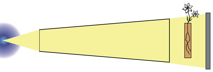
Figure 1 shows the configuration of a typical NR facility. NR requires a neutron source, which may be a reactor, the target of an elemental particle accelerator, or a neutron-emitting isotope. Thermal (12–100 meV) and cold (0.12–12 meV) neutrons are preferable for NR investigations. Therefore, epithermal, intermediate, and fast neutrons from the neutron source must be slowed图1显示了典型NR设施的配置。NR需要一个中子源,可以是反应堆、基本粒子加速器的靶或发射中子的同位素。热中子(12–100 meV)和冷中子(0.12–12 meV)更适用于NR研究。因此,来自中子源的超热中子、中能中子和快中子必须
Encyclopedia of Analytical Chemistry R.A. Meyers (Ed.) Copyright 2008 John Wiley & Sons Ltd down using a moderator such as heavy water. Neutrons enter a collimator that forms a neutron beam with specific geometric properties. The collimator may also contain filters that modify the energy spectrum of the beam or reduce the beam’s content of γ rays. The neutron beam is transmitted through the sample onto a plane position-sensitive detector, which is usually a scintillation screen. A charge-coupled device (CCD) camera coupled to the detector records a two-dimensional image that is a projection of the object on the detector plane. The advent of high-resolution digital cameras has allowed fast imaging and increased resolution. Digital imaging techniques that use a CCD camera combined with image-processing tools nowadays yield quantifiable images with a resolution of about 100 µm. Lehmann et al.(4) obtained a much higher spatial resolution with a special setup for microtomography. The required exposure time for such images is in the order of seconds. Images acquired using imaging plates or films (used until 1995) required several minutes of exposure to the potentially damaging neutron beam. The radiation dose received per image using modern techniques is about 0.003 mSv,(5) which is some two orders of magnitude less than the minimum value of 0.2 mSv h−1 found to affect plant growth.(6)
Encyclopedia of Analytical Chemistry R.A. Meyers (Ed.) Copyright 2008 John Wiley & Sons Ltd 利用重水等慢化剂进行减速。中子进入准直器,形成具有特定几何特性的中子束。准直器中还可装有过滤器,用以改变束流的能谱或降低束流中γ射线的含量。中子束穿过样品后投射到平面位置灵敏探测器上,该探测器通常为闪烁屏。与探测器耦合的电荷耦合器件(CCD)相机记录二维图像,即物体在探测器平面上的投影。高分辨率数码相机的出现实现了快速成像并提高了分辨率。如今,结合CCD相机与图像处理工具的数字成像技术可获得分辨率约为100 µm的可定量图像。Lehmann et al.(4) 利用专门的显微断层成像装置获得了更高的空间分辨率。此类图像所需的曝光时间为秒量级。使用成像板或胶片(沿用至1995年)获取图像则需要在具有潜在损伤作用的中子束下曝光数分钟。采用现代技术每幅图像所接受的辐射剂量约为0.003 mSv,(5) 比已知影响植物生长的最低剂量率0.2 mSv h−1约低两个数量级。(6)
3NEUTRON INTERACTIONS WITH THE PLANT–SOIL SYSTEM中子与植物–土壤系统的相互作用
NR is based on the Beer–Lambert exponential law of attenuation of radiation passing through matter(7):
NR基于辐射穿过物质时衰减所遵循的Beer–Lambert指数定律(7):

where I is the attenuated radiation (neutron) flux (cm−2 s−1), after an incident neutron flux I0 passes through a material of thickness d (cm) with an attenuating coefficient � (cm−1), which is a characteristic of the material. The attenuation coefficient, also called the macroscopic cross section, is related to the tabulated microscopic cross section σ (cm2) as with a nuclear density N (M). When a sample is placed in a neutron beam, heterogeneities in the composition and thickness of the sample result in variations in the intensity of the transmitted beam. Unlike X rays, neutron radiation interacts with atomic nuclei. There is no systematic change in the neutron attenuation coefficient with atomic number or mass. Each isotope has a specific neutron cross section, σ, which is also energy dependent. Hydrogen has a neutron cross section some 10 times greater than deuterium and also greater than many other elements in the soil–plant system.
式中,I为入射中子通量I0穿过厚度为d(cm)、衰减系数为�(cm−1)的材料后的衰减辐射(中子)通量(cm−2 s−1),衰减系数为该材料的特征参数。衰减系数又称宏观截面,它与列表给出的微观截面σ(cm2)及核密度N(M)相关。将样品置于中子束中时,样品组成和厚度的不均一性会导致透射束强度发生变化。与X射线不同,中子辐射与原子核发生相互作用。中子衰减系数并不随原子序数或原子质量呈系统性变化。每种同位素都具有特定的中子截面σ,且该截面还与能量有关。氢的中子截面约为氘的10倍,也大于土壤–植物系统中许多其他元素的中子截面。

NR reveals structures in plant–soil systems owing to differences in the � values of the system’s components. Table 1 shows a list of the chemical elements in the plant–soil system, along with their abundances and relative neutron cross sections. In both plant and soil, hydrogen is responsible for more than 90% of the neutron attenuation.
NR之所以能够显示植物–土壤系统中的结构,是由于系统各组分的�值存在差异。表1列出了植物–土壤系统中的化学元素及其丰度和相对中子截面。在植物和土壤中,氢均贡献了90%以上的中子衰减。
Some hydrogen is associated with organic molecules in the system; however, most hydrogen is water borne. Plant roots may thus be distinguished from soil due to their higher water content (θ). The gravimetric water content, θ (g g−1) of plant roots generally ranges between 0.7 and 0.95 g g−1, while that of soils at field capacity usually ranges between 0.12 and 0.3 g g−1. The structures that NR reveals in the soil–plant system are sensitive to θ.
系统中部分氢与有机分子结合,但大部分氢存在于水中。因此,植物根系可凭借其较高的含水量(θ)与土壤区分开来。植物根系的质量含水量θ(g g−1)一般为0.7–0.95 g g−1,而处于田间持水量的土壤通常为0.12–0.3 g g−1。NR所揭示的土壤–植物系统结构对θ十分敏感。
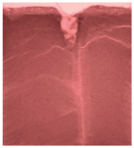
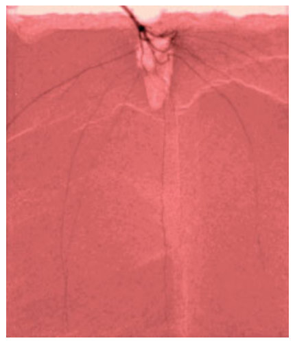
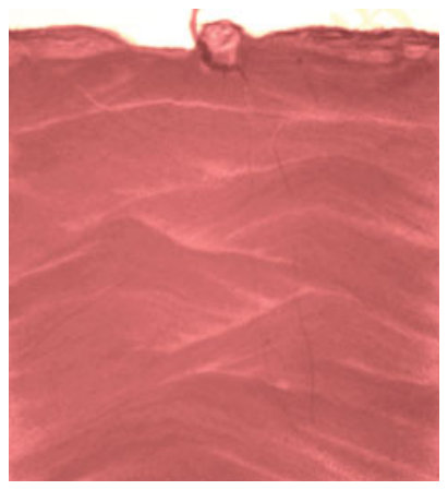
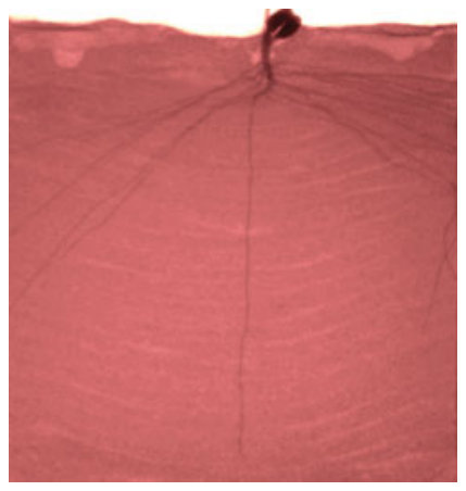
The high neutron attenuation coefficient of hydrogen is an important advantage of NR over X rays when applied to soil–plant system because the difference in water content allows the visualization of roots. The X-ray attenuation coefficients of root and soil components are less distinct (data not shown); therefore, the resulting radiograph has less contrast. Figure 2 shows that NR provides a better contrast between roots and soil than X rays.
应用于土壤–植物系统时,氢的高中子衰减系数是NR相对于X射线的重要优势,因为含水量的差异使根系得以显现。根系与土壤各组分的X射线衰减系数差异较小(数据未列出),因此所得照相图像的对比度较低。图2表明,NR在根系与土壤之间提供的对比度优于X射线。
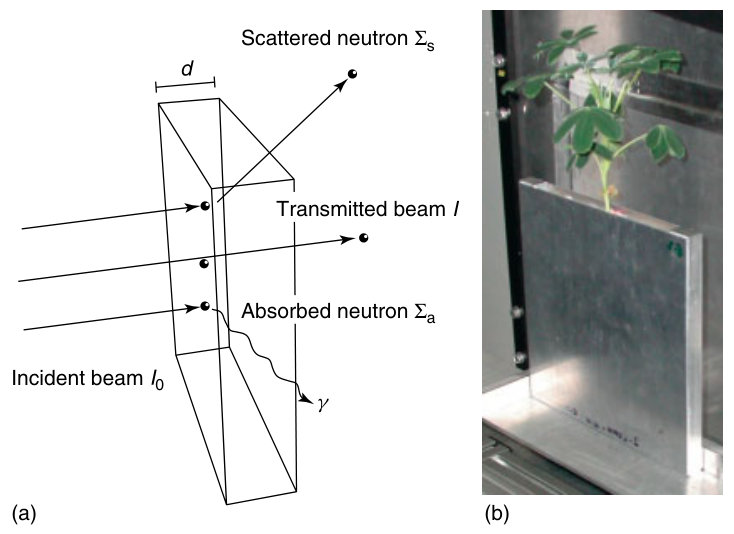
The attenuation coefficient (�) results from two types of neutron interactions with matter: absorption (�a) and scattering (�s) (Figure 3a). Hydrogen attenuates neutrons primarily by noncoherent elastic scattering.(11) Neutron scattering causes deviations from the exponential law of attenuation for thicker samples (more than a few millimeters) because some neutrons are multiple scattered into the detector plane, thus producing an artificially high signal. While the significance of neutron scattering is of little importance in the detection of roots in soil, it can be problematic when quantifying θ of the system. Algorithms such as the quantitative neutron imaging (QNI)(12) correct for the nonlinearity arising from neutron scattering. The QNI iteratively reconstructs the image by overlapping pointscattered functions calculated using a Monte Carlo simulation.
衰减系数(�)源自中子与物质的两类相互作用:吸收(�a)和散射(�s)(图3a)。氢主要通过非相干弹性散射使中子衰减。(11) 对于较厚的样品(超过几毫米),中子散射会导致偏离指数衰减定律,因为部分中子经多次散射后进入探测器平面,从而产生虚高的信号。虽然中子散射对检测土壤中的根系影响不大,但在定量系统的θ时可能造成问题。定量中子成像(QNI)(12)等算法可校正由中子散射引起的非线性。QNI通过叠加由Monte Carlo模拟计算得到的点散射函数来迭代重建图像。
NEUTRON RADIOGRAPHY FOR THE ANALYSIS OF PLANT–SOIL INTERACTIONS中子照相在植物–土壤相互作用分析中的应用
| Plant (mol kg−1)植物 (mol kg−1) | Soil (mol kg−1)土壤 (mol kg−1) | Element �(cm−1)元素 �(cm−1) | Plant neutron attenuation (cm−1)植物中子 衰减(cm−1) | Soil neutron attenuation (cm−1)土壤中子 衰减(cm−1) | |
|---|---|---|---|---|---|
| Hydrogen | 100 | 24 | 3.4 | 344 | 82 |
| Oxygen | 50 | 35 | 0.17 | 8.5 | 6 |
| Silicon | 71E–4 | 9.3 | 0.11 | _<0._1 | 1.02 |
| Carbon | 7.5 | 1.1 | 0.56 | 4.2 | 0.6 |
| Aluminum | 7.4E–4 | 2.1 | 0.1 | _<0._1 | 0.21 |
| Potassium | 5.1E–2 | 0.93 | 0.06 | _<0._1 | _<0._1 |
| Sodium | 8.7E–4 | 0.82 | 0.09 | _<0._1 | _<0._1 |
| Nitrogen | 0.21 | 3.6E–2 | 0.43 | _<0._1 | _<0._1 |
| Calcium | 2.5E–2 | 0.19 | 0.08 | _<0._1 | _<0._1 |
| Iron | 3.6E–4 | 0.19 | 1.2 | _<0._1 | 0.22 |
| Magnesium | 1.6E–2 | 7.8E–2 | 0.15 | _<0._1 | _<0._1 |
| Phosphorus | 1.3E–2 | 1.2E–2 | 0.12 | _<0._1 | _<0._1 |
| Titanium | 4.1E–6 | 2.4E–2 | 0.6 | _<0._1 | _<0._1 |
| Sulfur | 6.2E–3 | 2.3E–3 | 0.06 | _<0._1 | _<0._1 |
| Manganese | 1.8E–4 | 2.8E–3 | 1.2 | _<0._1 | _<0._1 |
| Chlorine | 5.6E–4 | 1.6E–3 | 1.3 | _<0._1 | _<0._1 |
| Zinc | 6.1E–5 | 5.3E–4 | 0.35 | _<0._1 | _<0._1 |
| Boron | 3.7E–4 | 1.8E–4 | 102 | _<0._1 | _<0._1 |
| Copper | 1.9E–5 | 1.6E–4 | 1.1 | _<0._1 | _<0._1 |
| Molybdenum | 2.1E–7 | 5.2E–5 | 0.52 | _<0._1 | _<0._1 |
| Total | 158 | 74 | 356 | 91 |
(a) (b)
4CONFIGURATION OF SOIL–PLANT SYSTEM FOR OPTIMAL IMAGING WITH NEUTRON RADIOGRAPHY适用于中子照相最佳成像的土壤–植物系统配置
A neutron radiograph is the result of all the neutron attenuation processes that occur when the neutron beam passes through a sample. The � value of the soil depends on θ and the neutron attenuation properties of its solid components. Soils that are high in iron or organic matter are unsuitable for NR because their high � values obscure the visualization of plant roots. The value of θ of the soil at the time of measurement should be as low as possible without inducing water stress in the plants. In practice, this represents the soil’s θ at a water potential of −1 bar. Silica sands have a low inherent � and a low θ at −1 bar. However, Menon et al.(5) showed that the high density and sharp edges of this material perturb normal root growth. Moradi et al. (unpublished data) tested a variety of plant growth media for their suitability in NR (Table 2).
中子照相图像是中子束穿过样品时发生的所有中子衰减过程的结果。土壤的�值取决于θ及其固相组分的中子衰减特性。富含铁或有机质的土壤不适用于NR,因为其较高的�值会妨碍植物根系的显现。测定时土壤的θ值应在不引起植物水分胁迫的前提下尽可能低。实际上,这相当于土壤在−1 bar水势下的θ。石英砂的固有�值较低,且在−1 bar时的θ也较低。然而,Menon et al.(5) 表明,该材料的高密度和锐利棱角会扰乱根系的正常生长。Moradi et al.(未发表数据)测试了多种植物生长基质对NR的适用性(表2)。
| Bulk density (g cm−3)堆积 密度 (g cm−3) | Water content at 1 bar (g g−1)1 bar时的 含水量 (g g−1) | �(dry material) (cm−1)�(干物质) (cm−1) | �(water content at −1 bar) (cm−1)�(−1 bar时 的含水量 下)(cm−1) | Notes注 | |
|---|---|---|---|---|---|
| Perlite | 0.125 | 1.08 | 0.1 | 4.5 | – |
| Porous glass beads | 0.49 | 0.16 | 1.6 | 2.5 | High_�_due to boron in glass |
| Ferrous | 1.4 | 0.08 | 1 | 1.6 | – |
| mine tailings | |||||
| Loam | 1.2 | 0.17 | 0.5 | 1.5 | – |
| Peat | 0.58 | 0.09 | 0.75 | 1.4 | – |
| Loamy sand | 1.3 | 0.09 | 0.3 | 0.75 | Normal root development |
| Fine quartz sand | 1.5 | 0.01 | 0.25 | 0.35 | Root develop- ment perturbed |
| Coarse quartz sand | 1.45 | 0.01 | 0.25 | 0.3 |
Table 2 shows that loamy sand, which permits normal root development, has a relatively low � at a θ at a water potential of −1 bar. Loamy sand has a higher θ at field表2表明,能保证根系正常发育的壤质砂土在−1 bar水势对应的θ下具有相对较低的�。壤质砂土在田间持水量下的θ
capacity (ca. 0.35 g g−1) than quartz sand (ca. 0.15 g g−1). Plants can thus be left longer in loamy sand without irrigation.
(约0.35 g g−1)高于石英砂(约0.15 g g−1)。因此,植物在壤质砂土中可以更长时间不灌溉。
Root visibility is proportional to root thickness, and inversely related to the width of the sample container.(13–17) The minimum detectable root thickness increases exponentially as the thickness of the soil profile increases. Moradi et al. (unpublished data) showed that using the loamy sand in Table 2, with a soil θ of 0.16 g g−1 (ca. −1 bar) and an average root θ of 0.85 g g−1, the minimum detectable root thickness R (mm) is related empirically to the slab thickness T (mm) according to the function:
根系的可见度与根的粗细成正比,而与样品容器的宽度成反比。(13–17) 最小可检测根径随土壤剖面厚度的增加呈指数增长。Moradi et al.(未发表数据)表明,使用表2中的壤质砂土,在土壤θ为0.16 g g−1(约−1 bar)、根系平均θ为0.85 g g−1的条件下,最小可检测根径R(mm)与土板厚度T(mm)之间的经验关系如下:

Roots are most easily visualized in thin containers. However, this condition is not conducive to plant growth since the walls of the container restrict root development. Therefore, one needs to find a balance between the ease of root visualization and the restriction of normal plant development.
在薄容器中最容易显现根系。然而,这一条件不利于植物生长,因为容器壁会限制根系发育。因此,需要在根系显现的难易程度与对植物正常发育的限制之间寻求平衡。
For many herbaceous species, a soil profile with a thickness of 12 mm provides enough space for relatively normal root development.(5) This gives a minimal detectable root thickness of 0.22 mm (Equation 2). Such a setup allows the visualization of the skeleton of the root system; however, fine roots (<2 mm in diameter) are undetectable.
对于许多草本物种而言,厚度为12 mm的土壤剖面可为根系提供相对正常发育的空间。(5) 此时最小可检测根径为0.22 mm(式2)。这种装置可以显现根系的骨架结构,但细根(直径<2 mm)无法检测。
5PRACTICAL CONSIDERATIONS实际操作注意事项
The sample container should be made of a material with a low �, suchas aluminum(� = 0.1 cm−1). Importantly, the system should not contain high concentrations of cobalt, which can form the persistent radioactive isotope Cobalt- 60, with a half-life of about five years, upon exposure to neutron radiation.
样品容器应由�值较低的材料制成,例如铝(� = 0.1 cm−1)。重要的是,系统中不应含有高浓度的钴,因为钴在中子辐照下会形成半衰期约为五年的持久性放射性同位素钴-60。
Filling the sample container with soil as homogeneously as possible provides better contrast for root visualization. Filling from the side of the container rather than from the top results in less structural heterogeneity associated with variations in the particle size distribution and thus pore size distribution, which are visible in the resulting neutron radiograph because of variations in soil water content. Figure 4 shows the effect of filling patterns caused by pouring soil from the top of the container.
尽可能均匀地向样品容器中填装土壤,可为根系显现提供更好的对比度。与从容器顶部填装相比,从容器侧面填装可减少因粒径分布及由此导致的孔隙大小分布变化而产生的结构不均一性;由于土壤含水量的差异,这种不均一性会在所得中子照相图像中显现出来。图4显示了从容器顶部倾倒土壤所形成的填装纹理的影响。
The suitability of NR to investigate root systems differs among plant species. As described earlier, thick roots with a high θ are more easily resolved than finer, drier roots. In a 10-mm-thick soil profile, the minimum detectable root size is ca. 0.16 mm (Equation 2). However, many plant species produce finer roots, which would not be visible in this system. Decreasing the thickness of the soil profile may permit the visualization of these roots, but may cause unnatural root growth patterns due to confinement. Moradi et al. (unpublished data) reported good root visibility in some members of the Fabaceae and Asteraceae families, whereas resolution was insufficient in Brassicacae, Solonaceae, and Poaceae. In principle, larger species such as small trees could be investigated using NR. However, this would require a larger container for nonperturbed growth. Consequently, the spatial resolution would decrease and one could only resolve large structural roots.
NR对根系研究的适用性因植物物种而异。如前所述,粗且θ高的根比细而干的根更容易分辨。在10 mm厚的土壤剖面中,最小可检测根径约为0.16 mm(式2)。然而,许多植物物种产生的根更细,在该系统中无法显现。减小土壤剖面厚度或可使这些根得以显现,但可能因空间受限而导致根系生长格局异常。Moradi et al.(未发表数据)报道,豆科(Fabaceae)和菊科(Asteraceae)部分物种的根系可见度良好,而十字花科(Brassicacae)、茄科(Solonaceae)和禾本科(Poaceae)的分辨率不足。原则上,NR也可用于研究小乔木等较大的物种,但这需要更大的容器以保证不受干扰的生长。因此,空间分辨率会降低,只能分辨较粗的结构根。
The water content of the soil and the plant should be monitored so that the soil water content is low enough at the time of imaging to provide adequate contrast yet not so low that the plant becomes water stressed. If a series of measurements are to be taken, then one can calculate the soil θ in the zone of interest from the results of each radiograph. This requires scattering correction and a water quantification algorithm calibrated for the particular plant–soil system. Water quantification using NR has the advantage over a gravimetric measurement because it can be used to determine the water content of the soil in the zone of interest, rather than providing an average value of a soil profile that may have a heterogeneous moisture distribution.
应监测土壤和植物的含水量,使成像时土壤含水量足够低以提供充分的对比度,但又不至于过低而使植物遭受水分胁迫。若需进行一系列测定,则可根据每幅照相图像的结果计算目标区域的土壤θ。这需要进行散射校正,并采用针对特定植物–土壤系统校准的水分定量算法。与称重法相比,利用NR定量水分的优势在于它能够测定目标区域的土壤含水量,而非仅给出水分分布可能不均一的土壤剖面的平均值。
The neutron beam formed in the collimator is not perfectly homogeneous. This also holds for the detector system. Therefore, there are spatial variations in intensity of the resulting radiography that are caused by the beam, rather than the sample. This requires that each radiograph be corrected by normalizing the image by a ‘‘flat field’’ or open-beam image with no sample. Similarly, noise generated by the camera assembly should be removed. A standard first step in image analysis is normalization, i.e. to transform recorded raw information relative values:
准直器中形成的中子束并非完全均匀,探测器系统亦是如此。因此,所得照相图像中存在由束流而非样品引起的强度空间变化。这就要求用无样品的“平场”或开束图像对每幅照相图像进行归一化校正。同样,相机组件产生的噪声也应予以去除。图像分析的标准第一步是归一化,即将记录的原始信息转换为相对值:
(b)(b)

where Iraw is the image as registered by the camera, Idark is the dark noise image without the beam, Iopenbeam contains the spatial field variation of the beam without object, and NORM is a factor to bring the resulting value into the valid range of the image-processing tools.
式中,Iraw为相机记录的图像, Idark为无中子束时的暗噪声图像,Iopenbeam包含无样品时中子束的空间场变化,NORM为使所得数值处于图像处理工具有效范围内的系数。
The background gray-level intensity within a neutron radiograph, caused, for example, by variations in the soil water content, may vary so much that a single global threshold cannot differentiate the roots satisfactorily. Menon et al.(5) overcame this problem by modifying an algorithm originally developed for resolving blood vessels in retinal images.(18)
中子照相图像中的背景灰度强度(例如由土壤含水量变化引起)可能变化很大,以至于单一的全局阈值无法令人满意地区分根系。Menon et al.(5) 通过改进一种最初用于分辨视网膜图像中血管的算法解决了这一问题。(18)
for example, may reveal species that avoid contaminant hotspots and reduce the risk of plant contaminant uptake. Similarly, understanding the mechanisms by which crop plants interact with patches of nutrients may aid the development of treatments to improve crop, and subsequently human, nutrition.
例如,可揭示哪些物种会避开污染热点,从而降低植物吸收污染物的风险。同样,了解农作物与养分斑块相互作用的机制,可能有助于开发改善作物营养进而改善人类营养的处理措施。
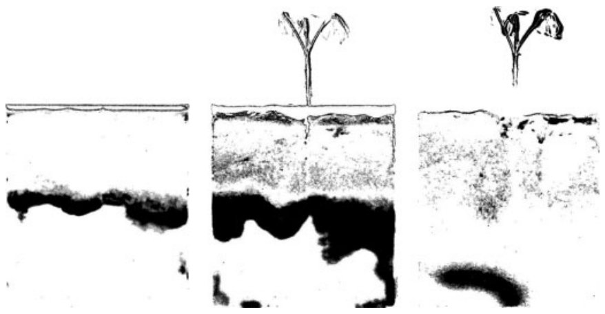
Perhaps more importantly, NR can quantify the spatial distribution and flux of water in the plant–soil system in near real time, in combination with root imaging. This permits the study of root water uptake and the effect of roots on the passage of water through soil. Figure 6 shows a series of images detailing water infiltration into a profile and water uptake by plant roots. Such studies have a wide variety of potential applications, such as selecting vegetation to minimize leaching from contaminated sites and the optimization of irrigation and fertilization systems.
或许更重要的是,NR能够结合根系成像,近实时地定量植物–土壤系统中水分的空间分布和通量。这使得研究根系吸水以及根系对水分在土壤中运移的影响成为可能。图6展示了一系列图像,详细描绘了水分向剖面中的入渗以及植物根系对水分的吸收。此类研究具有广泛的潜在应用,例如选择植被以最大限度减少污染场地的淋溶,以及优化灌溉和施肥系统。
7CONCLUSIONS AND OUTLOOK结论与展望
6APPLICATION应用
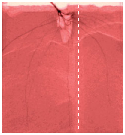
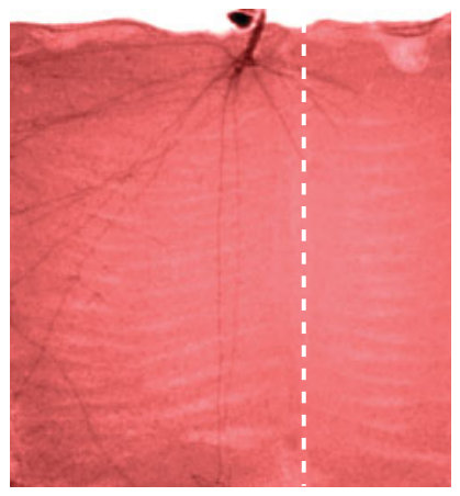
The application of NR can enhance the study of root development and root–soil interactions by revealing the location of roots over time without disturbance. Plant root development is a function of the plant species and the nature of the soil into which they penetrate. Soil components, such as organic matter, nutrients, and contaminants, occur heterogeneously. NR is an ideal tool to study how roots interact with patches of low or high concentration. When roots encounter a patch or discontinuity in soil, they may proliferate, wither, or continue growing unaffected. Figure 5(a) and (b) shows how NR can reveal plant responses to a patch of nickel, a toxic heavy metal, in soil. Such experiments,
NR能够在不干扰的情况下揭示根系随时间的位置变化,因而可促进对根系发育及根–土相互作用的研究。植物根系的发育取决于植物物种及其所穿透土壤的性质。土壤组分(如有机质、养分和污染物)呈不均匀分布。NR是研究根系如何与低浓度或高浓度斑块相互作用的理想工具。当根系遇到土壤中的斑块或不连续界面时,可能会增殖、枯萎或不受影响地继续生长。图5(a)和(b)展示了NR如何揭示植物对土壤中镍(一种有毒重金属)斑块的响应。此类试验,
The quantification of root mass and soil water content using NR of plant–soil interactions has considerable scope for improvement, particularly, the processing of raw images obtained at the neutron facility. Refinement of root segmentation algorithms described by Menon et al.(5) would greatly enhance the accuracy and precision of the technique. One obvious drawback of NR is that, at present, it requires access to a specialized facility, of which there are only a few available worldwide with the desirable performance. Competition for beam time is fierce, since NR finds applications in many fields of science. However, the usefulness of NR indicates that it may follow the same pattern of development as synchrotron radiation facilities. Initially, financial and technical constraints limited their application, but their usefulness ensured subsequent technological development and capital injection, and thus they became commonplace and the standard equipment for some analyses.
利用NR研究植物–土壤相互作用以定量根系生物量和土壤含水量的方法仍有很大的改进空间,特别是对在中子设施获得的原始图像的处理。改进Menon et al.(5) 所述的根系分割算法将大大提高该技术的准确度和精密度。NR的一个明显缺点是目前需要使用专门的设施,而全球具备理想性能的此类设施屈指可数。由于NR在众多科学领域均有应用,束流时间的竞争十分激烈。然而,NR的实用价值表明,它可能会遵循与同步辐射设施相同的发展模式。同步辐射设施最初受资金和技术条件限制而应用有限,但其实用价值确保了后续的技术发展和资金投入,从而使其得以普及,并成为某些分析的标准设备。
(b)(b)
NEUTRON RADIOGRAPHY FOR THE ANALYSIS OF PLANT–SOIL INTERACTIONS
中子照相在植物–土壤相互作用分析中的应用
(a)
(b) (c)
RELATED ARTICLES相关文章
Nuclear Methods (Volume 14)核方法(第14卷)
Nuclear Reaction Analysis • Prompt γ -Neutron Activation Analysis • Scattering and Absorption of γ -Rays and Thermalization and Disappearance of Neutrons
核反应分析 • 瞬发γ中子活化分析 • γ射线的散射与吸收以及中子的热化与消失
Environment: Water and Waste (Volume 4)环境:水与废弃物(第4卷)
Neutron Activation in Environmental Analysis • Soil Instrumental Methods
环境分析中的中子活化 • 土壤仪器分析方法
REFERENCES参考文献
1. R.O. Kuchenbuch, K.T. Ingram, ‘Image Analysis for Nondestructive and Non-invasive Quantification of Root Growth and Soil Water Content in Rhizotrons’, J. Plant Nutr. Soil Sc., 165(5), 573–581 (2002).
2. P.J. Gregory, D.J. Hutchison, D.B. Read, P.M. Jenneson, W.B. Gilboy, E.J. Morton, ‘Non-invasive Imaging of Roots with High Resolution X-ray Micro-tomography’, Plant Soil, 255(1), 351–359 (2003).
3. S.T. Willatt, R.G. Struss, H.M. Taylor, ‘In situ Root Studies Using Neutron Radiography’, Agron. J., 70(4), 581–586 (1978).
4. E.H. Lehmann, G. Frei, G. Kuhne, P. Boillat, ‘The Microsetup for Neutron Imaging: A Major Step Forward to
Improve the Spatial Resolution’, Nucl. Instrum. Methods A, 576(2–3), 389–396 (2007).
5. M. Menon, B. Robinson, S.E. Oswald, A. Kaestner, K.C. Abbaspour, E. Lehmann, R. Schulin, ‘Visualization of Root Growth in Heterogeneously Contaminated Soil Using Neutron Radiography’, Eur. J. Soil Sci., 58(3), 802–810 (2007).
6. A. Real, S. Sundell-Bergman, J.F. Knowles, D.S. Woodhead, I. Zinger, ‘Effects of Ionizing Radiation Exposure on Plants, Fish and Mammals: Relevant Data for Environmental Radiation Protection’, J. Radiol. Prot., 24(4A), A123–A137 (2004).
7. S. Kasperl, P. Vontobel, ‘Application of an Iterative Artefact Reduction Method to Neutron Tomography’, Nucl. Instrum. Meth. A, 542(1–3), 392–398 (2005).
8. F.B. Salisbury, C.W. Ross, Plant Physiology, Wadsworth, Belmont, CA, 1992.
9. B. Mason, C. Moore, Principles of Geochemistry, John Wiley & Sons, Hong Kong, 1982.
10. PSI (2008) ‘Comparison Between Neutron and X-ray Attenuation’, from http://neutra.web.psi.ch/images/diag x n e.gif.
11. D.G. Allis, B.S. Hudson, ‘Inelastic Neutron Scattering Spectrum of Cs-2[B12H12]: Reproduction of its Solid-state Vibrational Spectrum by Periodic DFT’, J. Phys. Chem. A, 110(10), 3744–3749 (2006).
12. R.K. Hassanein, Correction Methods for the Quantitative Evaluation of Thermal Neutron Tomography, ETH Zurich, Switzerland, 2006.
13. T.M. Nakanishi, M. Matsubayashi, ‘Nondestructive Water Imaging by Neutron Beam Analysis in Living Plants’, J. Plant Physiol., 151(4), 442–445 (1997).
14. J. Furukawa, T.M. Nakanishi, H. Matsubayashi, ‘Neutron Radiography of a Root Growing in Soil with Vanadium’, Nucl. Instrum. Meth. A, 424(1), 116–121 (1999).
15. T.M. Nakanishi, Y. Okuni, J. Furukawa, K. Tanoi, H. Yokota, N. Ikeue, M. Matsubayashi, H. Uchida, A. Tsiji, ‘Water Movement in a Plant Sample by Neutron Beam Analysis as well as Positron Emission Tracer Imaging System’, J. Radioanal. Nucl. Chem., 255(1), 149–153 (2003).
16. U. Matsushima, Y. Kawabata, M. Hino, P. Geltenbort, B.M. Nicolai, ‘Measurement of Changes in Water
Thickness in Plant Materials Using Very Low-energy Neutron Radiography’, Nucl. Instrum. Meth. A, 542(1–3), 76–80 (2005).
17. T.M. Nakanishi, Y. Okuni, Y. Hayashi, H. Nishiyama, ‘Water Gradient Profiles at Bean Plant Roots Determined by Neutron Beam Analysis’, J. Radioanal. Nucl. Chem., 264(2), 313–317 (2005).
18. A. Hoover, V. Kouznetsova, M. Goldbaum, ‘Locating Blood Vessels in Retinal Images by Piecewise Threshold Probing of a Matched Filter Response’, IEEE Trans. Med. Imag., 19(3), 203–210 (2000).