REGULAR ARTICLE
Ahmad B. Moradi & Héctor M. Conesa & Brett Robinson & Eberhard Lehmann & Guido Kuehne & Anders Kaestner & Sascha Oswald & Rainer Schulin
Received: 8 August 2008 /Accepted: 10 November 2008 /Published online: 9 December 2008 # Springer Science + Business Media B.V. 2008
Abstract Neutron Radiography (NR) is a valuable non-invasive tool to study in situ root development in soil. However, there is a lacuna of quantitative information on its capabilities and limitations. We combined neutron radiography with image analysis techniques to quantify the neutron absorption coefficients (Σ) of various root-growth media for a range of water contents (θ) in the presence and absence of plant roots with various rooting systems. Plants were grown in aluminium containers (170×150×12 mm) and were imaged using NR, as well as X-Ray radiography and an optical scanner. Sandy soil was the best medium for NR because it supported plant growth at θ that gave a good contrast for root visualisation. After correction for neutron scattering, we obtained a linear correlation between Σ and soil θ. The minimum detectable root thickness in neutron radiographs was found to be 0.2 mm in these containers. Combining NR with X-Ray radiography could provide information on soil structure in addition to revealing root structure and development.
摘要 中子成像(Neutron Radiography,NR)是原位研究土壤中根系发育的一种有价值的非侵入性工具。然而,有关其性能与局限性的定量信息尚属空白。本研究将中子成像与图像分析技术相结合,定量测定了多种根系生长介质在不同含水量(θ)条件下、在有无不同根系类型植物根系存在时的中子吸收系数(Σ)。植物生长于铝制容器(170×150×12 mm)中,并采用NR、X射线成像及光学扫描仪进行成像。砂质土壤是最适合NR的介质,因其在可为根系显像提供良好对比度的θ条件下仍能支持植物生长。经中子散射校正后,Σ与土壤θ之间呈线性相关。在本研究所用容器中,中子成像图像中可检测的最小根系粗度为0.2 mm。将NR与X射线成像相结合,除揭示根系结构与发育外,还可提供土壤结构信息。
Keywords Neutron radiography. Neutron attenuation coefficient . Neutron scattering . Plant roots . Soil water content . X-ray radiography
关键词 中子成像;中子衰减系数;中子散射;植物根系;土壤含水量;X射线成像
1Introduction引言
Responsible Editor: Hans Lambers. A. B. Moradi (*) : H. M. Conesa : B. Robinson : R. Schulin Institute of Terrestrial Ecosystems, Soil Protection group, ETH Zurich, CHN F28.2, Universitaetstrasse 16, CH-8092 Zurich, Switzerland e-mail: ahmad.moradi@env.ethz.ch URL: http://www.ito.ethz.ch/soilprot/
E. Lehmann : G. Kuehne : A. Kaestner Paul Scherrer Institut, Villigen, Switzerland S. Oswald Helmholtz Centre for Environmental Research - UFZ, Leipzig, Germany
E. Lehmann : G. Kuehne : A. Kaestner 保罗·谢勒研究所(Paul Scherrer Institut),瑞士 Villigen S. Oswald 亥姆霍兹环境研究中心(Helmholtz Centre for Environmental Research - UFZ),德国莱比锡
Neutron Radiography (NR) is one of the few non-destructive techniques available to image living plant roots in situ (Menon et al. 2007). Roots grow in a heterogeneous, porous, semi-compressible medium compound of solid, liquid and gaseous phases, known as soil. It is difficult to observe tightly enmeshed roots in an opaque soil matrix without disturbance. Better understanding of root behaviour in heterogeneous soil, requires efficient non-invasive techniques to analyze root and soil structure in situ (Fitz et al. 2005; Pierret et al. 2005; Nakaji et al. 2008; Pierret 2008).
中子成像(NR)是少数几种可对活体植物根系进行原位成像的无损技术之一(Menon et al. 2007)。根系生长于由固、液、气三相组成的非均质、多孔、半可压缩介质——即土壤之中。在不产生扰动的情况下,很难观察到紧密交织于不透明土壤基质中的根系。要更好地理解根系在非均质土壤中的行为,需要借助高效的非侵入性技术对根系和土壤结构进行原位分析(Fitz et al. 2005; Pierret et al. 2005; Nakaji et al. 2008; Pierret 2008)。
Current non-invasive and non-destructive methods for studying roots include minirhizotrons which are transparent plastic tubes inserted into the ground to view the roots using a camera (Kuchenbuch and Ingram 2002), magnetic resonance imaging (Bottomley et al. 1993; Doussan et al. 2003; Heyes and Clark 2003), neutron radiography and tomography (Willatt et al. 1978; Willatt and Struss 1979; Bois and Couchat 1983; Nakanishi et al. 2005; Menon et al. 2007; Oswald et al. 2008; Tumlinson et al. 2008) as well as X-Ray imaging and tomography (Moran et al. 2000; Naftel et al. 2001; Gregory et al. 2003; Pierret et al. 2003b). Root observation using minirhizotrons is non-destructive. However, they have an invasive component that may perturb root development and water flux (Majdi 1996). Ferromagnetic materials hinder magnetic resonance imaging (Hall et al. 1997), thus limiting this technique to carefully selected media such as pretreated sand and soil, agar, and glass beads. NR provides images similar to X-Ray radiography. Neutrons interact with the nuclei of atoms while X-Rays interact with electrons. Whereas X-Ray attenuation depends on element’s atomic number Z, the neutron attenuation coefficient is independent of Z and only a few elements such as hydrogen strongly attenuate neutrons. Therefore, H-rich, organic materials and water are clearly visible in neutron radiographs, while many soil components and structural materials such as Si, Ca and Al are nearly transparent. This permits in situ investigations of specimens housed in various types of experimental apparatus. Such housings often impair or prevent X-Ray imaging because of their high X-Ray cross sections. However the neutron cross section of these elements is moderately low, allowing the neutrons to pass through undisturbed.
目前用于研究根系的非侵入性和无损方法包括:微根管法,即将透明塑料管插入地下并用摄像头观察根系(Kuchenbuch and Ingram 2002);磁共振成像(Bottomley et al. 1993; Doussan et al. 2003; Heyes and Clark 2003);中子成像及断层扫描(Willatt et al. 1978; Willatt and Struss 1979; Bois and Couchat 1983; Nakanishi et al. 2005; Menon et al. 2007; Oswald et al. 2008; Tumlinson et al. 2008);以及X射线成像及断层扫描(Moran et al. 2000; Naftel et al. 2001; Gregory et al. 2003; Pierret et al. 2003b)。利用微根管观察根系是无损的,但其具有一定的侵入性,可能干扰根系发育和水分通量(Majdi 1996)。铁磁性物质会妨碍磁共振成像(Hall et al. 1997),因而该技术仅限于经过精心选择的介质,如经预处理的砂和土壤、琼脂以及玻璃珠。NR所得图像与X射线成像相似。中子与原子核相互作用,而X射线则与电子相互作用。X射线的衰减取决于元素的原子序数Z,而中子衰减系数与Z无关,仅有少数元素(如氢)对中子有强烈衰减作用。因此,富氢的有机物质和水在中子成像图像中清晰可见,而许多土壤组分及结构材料(如Si、Ca和Al)则几乎是透明的。这使得对置于各类实验装置中的样品进行原位研究成为可能。此类外壳由于X射线截面较高,常会削弱甚至阻碍X射线成像;而这些元素的中子截面则相对较低,使中子能够不受干扰地穿过。
NR is a valuable tool for studying root behaviour in soil since the difference in gravimetric water content (θ) of roots and the surrounding soil provides enough contrast to visualize the roots. Nakanishi et al. (2005) showed how soil θ changes along a vertical distance from the root surface using thermal neutron beam analysis. Microscopic movement of water around the roots of soybean seedlings was studied by Furukawa et al. (1999). Menon et al. (2007) visualized the root growth of lupin plants over a period of 3 weeks and revealed how heterogeneous soil can affect root growth. Fine roots (<2 mm) are particularly important for the uptake of water and nutrient by plants (Sutton and Tinus 1983; Waisel et al. 2002). While it is widely recognized that the total length of the fine roots is generally larger than that of the coarse roots of a plant, it is also acknowledged that the amount of fine roots is often underestimated because of the lack of reliable measurement techniques, their small size and near transparency (Vogt et al. 1998; Costa et al. 2001; Pierret et al. 2003a). Root visibility in NR is proportional to root thickness and inversely proportional to soil moisture content (Furukawa et al. 1999; Nakanishi et al. 2003, 2005; Matsushima et al. 2005; Menon et al. 2007). These authors used either thin containers (as thin as 2 mm) or artificial plant growth media (quartz sand or silt). There is a lacuna of information on the performance of NR in other systems, for example with topsoils, and more realistic growing containers. The effects of root thickness, soil type and the water content of the surrounding soil on the contrast between soil and roots in NR images has not been quantified and the capability of NR has not yet been evaluated in visualizing fine roots with various thickness in various soil types and water contents.
NR是研究土壤中根系行为的有价值工具,因为根系与周围土壤在质量含水量(θ)上的差异可提供足够的对比度以使根系显像。Nakanishi et al.(2005)利用热中子束分析揭示了土壤θ沿垂直于根表面方向随距离的变化。Furukawa et al.(1999)研究了大豆幼苗根系周围水分的微观运移。Menon et al.(2007)对羽扇豆植株3周内的根系生长进行了可视化,揭示了非均质土壤对根系生长的影响。细根(<2 mm)对植物吸收水分和养分尤为重要(Sutton and Tinus 1983; Waisel et al. 2002)。人们普遍认识到,植物细根的总长度通常大于粗根,但同时也承认,由于缺乏可靠的测定技术,加之细根尺寸小且近乎透明,细根数量常被低估(Vogt et al. 1998; Costa et al. 2001; Pierret et al. 2003a)。在NR中,根系的可见度与根系粗度成正比,与土壤含水量成反比(Furukawa et al. 1999; Nakanishi et al. 2003, 2005; Matsushima et al. 2005; Menon et al. 2007)。上述研究者或使用薄型容器(薄至2 mm),或使用人工植物生长介质(石英砂或粉砂)。关于NR在其他体系(例如表层土壤及更接近实际的栽培容器)中的表现,目前尚缺乏相关信息。根系粗度、土壤类型及周围土壤含水量对NR图像中土壤与根系之间对比度的影响尚未得到定量,NR在不同土壤类型和含水量条件下对不同粗度细根的显像能力也尚未得到评估。
The quantitative analysis of a neutron radiograph requires corrections for interactions between neutron beam and the sample other than adsorption, which can be significant. These types of interactions can be coherent, incoherent, elastic an inelastic scattering. Calibrating of the θ against neutron transmission usually may not give a satisfactory result, especially at higher θ where transmission decreases and multiple neutron scattering becomes important leading to a considerable deviation from the exponential law of attenuation (Hassanein 2006). While neutron scattering is used to measure various properties of materials (Bailey 2003), it needs to be corrected for studying water and hydrogen rich organic materials, such as roots, in soil.
对中子成像图像进行定量分析时,需要对中子束与样品之间除吸收以外的相互作用进行校正,这些作用可能相当显著。此类相互作用包括相干散射、非相干散射、弹性散射和非弹性散射。将θ与中子透射率进行校准通常难以获得满意的结果,尤其是在θ较高时,透射率下降,多重中子散射变得重要,从而导致其明显偏离指数衰减定律(Hassanein 2006)。尽管中子散射可用于测定材料的多种性质(Bailey 2003),但在研究土壤中的水分及富氢有机物质(如根系)时,必须对其加以校正。
We aimed to assess the capabilities and limitations of NR combined with image analysis tools for studying non-destructively root development in soil with particular attention to the quantitative effects of neutron scattering, soil type, soil θ and root thickness on the root visibility in soil. Specifically, we sought to quantify the effects of soil type, soil moisture, and container dimensions and size on the ability to visualise roots and determine the detection limits for NR under realistic conditions for plant growth. Furthermore, we aimed to develop NR as a tool to quantify the water content of soil in the rhizosphere.
本研究旨在评估NR结合图像分析工具无损研究土壤中根系发育的能力与局限性,并特别关注中子散射、土壤类型、土壤θ及根系粗度对土壤中根系可见度的定量影响。具体而言,我们试图定量分析土壤类型、土壤含水量以及容器尺寸和大小对根系显像能力的影响,并确定在接近实际植物生长条件下NR的检测限。此外,我们还旨在将NR发展为定量测定根际土壤含水量的工具。
2Materials and methods材料与方法
3Neutron and X-Ray radiography systems中子与X射线成像系统
The Beer-Lambert law describes the attenuation of both neutron and X-Ray radiation (Kasperl and Vontobel 2005), assuming that the neutron detector records only the uncollided flux component and that the neutrons are mono-energetic or have a small wavelength bandwidth:
假设中子探测器仅记录未碰撞的通量分量,且中子为单能或波长带宽较窄,则中子和X射线辐射的衰减均可用Beer-Lambert定律描述(Kasperl and Vontobel 2005):

where I is the detected neutron flux (cm−2 s−1) after an incident neutron flux, I0, passes through material of thickness d (cm) with a neutron attenuation coefficient of Σ (cm−1), which is a bulk physical property of the material. The material may be crystalline, polymorphic, or, as in our case, a porous media.
式中,I为入射中子通量I0穿过厚度为d(cm)、中子衰减系数为Σ(cm−1)的材料后被探测到的中子通量(cm−2 s−1),Σ是材料的一种整体物理性质。该材料可以是晶体、多晶型物质,或如本研究中的多孔介质。
The experiments were performed at the neutron and X-Ray radiography facilities at Paul Scherrer Institut (PSI), Villigen, Switzerland. Both neutron imaging facilities at PSI; NEUTRA for thermal neutrons (Lehmann et al. 1999), and ICON for imaging with cold neutrons (Kuhne et al. 2005), consist of the neutron source (the spallation neutron source SINQ), a special collimator, and a twodimensional image detector. For a thermal spectrum, the moderator is kept at about room temperature (∼300 K), which yields neutron energies of around 25 meV and velocities of around 2200 m/s. For a cold spectrum, liquid deuterium is used as an additional moderator at around 25° K, yielding neutron energies of around 5.7 meV and velocities of about 1000 m/s. The collimator is a beam-forming assembly that determines the geometric properties of the beam and contains filters that reduce the intensity of accompanying gamma rays. The image resolution depends on the collimator geometry and is expressed by the collimation ratio L/D, where L is the collimator length and D is the diameter of the inlet aperture of the collimator on the side facing the source. In our study, a neutron flux of 4.2×106 cm−2 s−1 with a mean energy of 25 meV and a collimation ratio of L/D= 550, was used for NEUTRA while for ICON a neutron flux of 2.8×106 cm−2 s−1, a mean energy of 5.7 meV, and a collimation ratio of L/D=605. The beam was transmitted through the sample and an area position-sensitive detector recorded the resulting image, which is integrated in the beam direction over the thickness, d. This detector records a two-dimensional image that is a projection of the object on the detector plane. We used a CCD camera detector with an array of 1024×1024 pixels in conjunction with a neutron-sensitive 6Li based scintillator screen (Applied scintillation technologies, UK), giving a resolution of 110–170 μm in the digital images.
实验在瑞士Villigen保罗·谢勒研究所(PSI)的中子和X射线成像设施上进行。PSI的两套中子成像设施——用于热中子的NEUTRA(Lehmann et al. 1999)和用于冷中子成像的ICON(Kuhne et al. 2005)——均由中子源(散裂中子源SINQ)、专用准直器和二维图像探测器组成。对于热中子谱,慢化剂保持在约室温(∼300 K),所产生的中子能量约为25 meV,速度约为2200 m/s。对于冷中子谱,则以约25° K的液态氘作为附加慢化剂,所产生的中子能量约为5.7 meV,速度约为1000 m/s。准直器是决定射束几何特性的束流成形组件,其中装有可降低伴生γ射线强度的滤片。图像分辨率取决于准直器几何结构,以准直比L/D表示,其中L为准直器长度,D为准直器面向中子源一侧入口孔径的直径。本研究中,NEUTRA采用的中子通量为4.2×106 cm−2 s−1,平均能量为25 meV,准直比L/D= 550;ICON采用的中子通量为2.8×106 cm−2 s−1,平均能量为5.7 meV,准直比L/D=605。射束穿过样品后,由面位置灵敏探测器记录所得图像,该图像沿射束方向在厚度d上积分。探测器记录的二维图像为物体在探测器平面上的投影。我们使用具有1024×1024像素阵列的CCD相机探测器,并配合基于6Li的中子敏感闪烁屏(Applied scintillation technologies,英国),数字图像分辨率为110–170 μm。
The X-Ray radiography facility at PSI consists of an X-Ray tube with a maximal voltage of 320 kV. Using the focal spot at the tungsten target of about 3 mm, which corresponds to arrangement of the tube just in front of the outer collimator close to the target block wall, we obtained about the same L/D ratio as with the neutron beam. A scintillator screen made of Gadolinium-oxy-sulfide (Gadox) was used for X-Ray imaging.
PSI的X射线成像设施由最高电压为320 kV的X射线管组成。使用钨靶上约3 mm的焦斑(对应于将X射线管布置在靠近靶块壁的外准直器正前方),所获得的L/D比与中子束大致相同。X射线成像采用硫氧化钆(Gadox)制成的闪烁屏。
4Materials testing材料测试
We chose a variety of materials that could be potentially used as root growth media (Table 1). The materials were air-dried and packed into the plant growth containers and were imaged the same way as the plant roots. We used containers with inner dimensions of 0.17×0.15×0.012 m. The containers were made of aluminium because of its low neutron attenuation and therefore high transparency.
我们选择了多种可能用作根系生长介质的材料(表1)。将这些材料风干后装入植物生长容器,并以与植物根系相同的方式成像。所用容器内部尺寸为0.17×0.15×0.012 m。容器由铝制成,因为铝的中子衰减低,因而透明度高。
5Calibration for soil water content土壤含水量校准
The NR image is a projection of the object (in our case a container with a thickness of 12 mm) in the direction of the container thickness. Thus both soil water and water residing in the roots contribute to the neutron attenuation coefficients calculated pixel-wise in each image. The contrast in the NR images comes from difference in water content of the roots and the surrounding soil. Calibrating neutron attenuation coefficients against soil water is needed to calculate the contribution of root water to the Σ, knowing the root diameter. To calibrate neutron transmission and Σ against soil θ, three containers were filled with the sandy soil (Table 1) in the same way as described above. The soils were saturated with water, then left to dry for 2 weeks at room temperature by opening one side of the container. The containers were imaged seven times during the drying period. Before each imaging, the total water content of the soil was measured gravimetrically.
NR图像是物体(本研究中为厚度12 mm的容器)沿容器厚度方向的投影。因此,土壤水和根系中的水均对每幅图像中逐像素计算的中子衰减系数有贡献。NR图像中的对比度来源于根系与周围土壤含水量的差异。在已知根系直径的情况下,需要将中子衰减系数与土壤水分进行校准,以计算根系水分对Σ的贡献。为将中子透射率和Σ与土壤θ进行校准,按上述相同方法将砂质土壤(表1)装入三个容器中。土壤经水饱和后,打开容器的一侧,在室温下干燥2周。在干燥期间对容器进行了七次成像。每次成像前,采用称重法测定土壤的总含水量。
| Particle size (mm)粒径 (mm) | Bulk density (g cm−3)容重 (g cm−3) | Porosity (% v/v)孔隙度 (% v/v) | Water holding capacity at 100 kPa (cm3/cm−3)100 kPa下的持水量 (cm3/cm−3) | Provenience来源 | |
|---|---|---|---|---|---|
| Perlite | <2 | 0.125 | 97 | 0.91 | Samen Mauser, Switzerland |
| Porous glass beads | 1.5–2.5 | 0.49 | 71 | 0.16 | Schott, Germany |
| Mine tailing soil | <2 | 1.4 | 35 | 0.08 | Southeast, Spain |
| Loamy soil | <2 | 1.2 | 49 | 0.17 | Mattenweg, Switzerland |
| Organic soil | <2 | 0.58 | 88 | 0.09 | Migros, Switzerland |
| Sandy soil | <2 | 1.3 | 36 | 0.09 | Eiken, Switzerland |
| Fine quartz sand | 0.1–0.7 | 1.5 | 19 | 0.01 | Carlo, Switzerland |
| Coarse quartz sand | 0.7–1.5 | 1.45 | 26 | 0.01 | Carlo, Switzerland |
6Plant growth植物培养
7Imaging procedure成像步骤
Three plant species, with three different rooting systems were selected for this study; garden tomato (Lycopersicon esculentum Mill.) with a fibrous root system of fine roots, chickpea (Cicer arietinum L.) with strong tap root system and Berkheya coddii Roessl with a fibrous root system of both fine and coarse roots. We filled 12 containers with the sandy loam described in Table 1. There were four treatments, each with three replicates: the three aforementioned plants and an unplanted treatment. The average bulk density of the packed soil was 1.3 g cm−3. The seeds of the plants were sown directly onto the soil surface at the centre of the containers except for Berkheya coddii, where seedlings were transplanted into the containers. The plants were grown for 7 weeks before the imaging in a controlled environment chamber at 16–19°C and a daily photoperiod of 16 h generated by fluorescent lighting (0.4–0.6 lumen cm−3). Plants were irrigated with Hoagland’s nutrient solution (Hoagland and Arnon 1938) to maintain soil θ of around 20%. Irrigation was stopped 3 days before imaging. Neutron and X-Ray radiographs were taken on the same day. After imaging, we opened one side of the containers and scanned the soil surface using a conventional photo scanner with a pixel array of 2250×2250. Soil in the containers was subdivided into sections. Each section was weighted and the roots were carefully separated from the soil. The dry mass of the roots as well as the moisture content of the soil in each section were determined by weighing.
本研究选取了具有三种不同根系类型的三种植物:具有细根须根系的番茄(Lycopersicon esculentum Mill.)、具有强壮直根系的鹰嘴豆(Cicer arietinum L.),以及兼具细根和粗根须根系的Berkheya coddii Roessl。将表1所述砂壤土装入12个容器中。共设四个处理,每个处理三个重复:上述三种植物及一个不种植处理。装填土壤的平均容重为1.3 g cm−3。除Berkheya coddii为移栽幼苗外,其余植物种子均直接播于容器中央的土壤表面。植物在人工气候室中生长7周后进行成像,温度为16–19°C,由荧光灯提供每日16 h的光周期(0.4–0.6 lumen cm−3)。植物以Hoagland营养液(Hoagland and Arnon 1938)灌溉,使土壤θ保持在20%左右。成像前3天停止灌溉。中子和X射线成像在同一天进行。成像后,打开容器的一侧,使用像素阵列为2250×2250的常规照片扫描仪扫描土壤表面。将容器中的土壤分成若干区段,逐段称重,并小心地将根系从土壤中分离。通过称重测定各区段根系干重及土壤含水量。
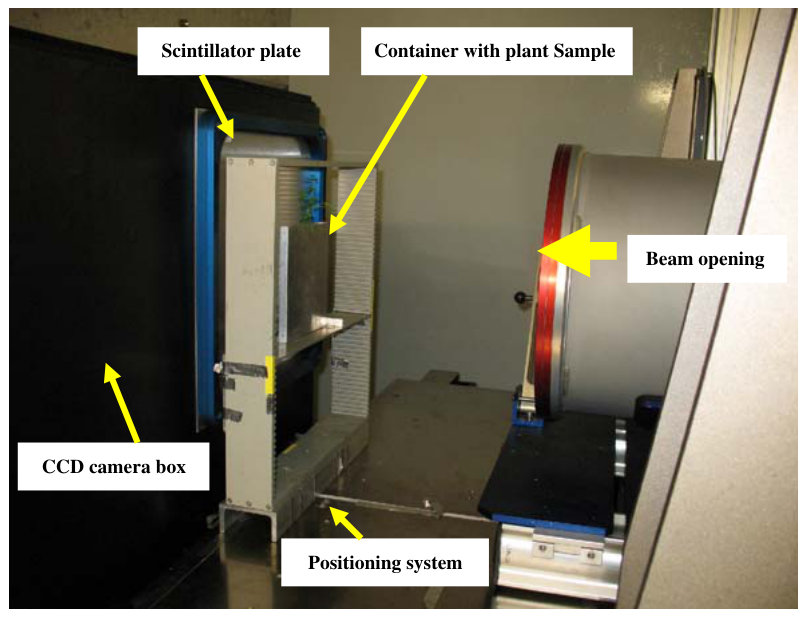
All containers were imaged in exactly the same position using an automated positioning table in front of the beam line with a distance of 10 cm from the scintillator plane in the ICON setup and 15 cm in the NEUTRA and X-Ray setups (Fig. 1). We used exposure times of 45, 15 and 10 s for ICON, NEUTRA and X-Ray images respectively. These exposure times gave the best signal to noise ratio and therefore the best contrast between the roots and the soil. X-Ray images used a voltage of 120 kV at 10 mA. To correct for spatial variations in beam intensity, we collected open beam images and dark current images. The field of view was 19×17 cm which left a margin (area without sample) of 1 cm. This was used to normalise the images and to correct for any fluctuations in beam intensity.
所有容器均利用射束线前方的自动定位台在完全相同的位置成像,在ICON装置中与闪烁屏平面的距离为10 cm,在NEUTRA和X射线装置中为15 cm(图1)。ICON、NEUTRA和X射线图像的曝光时间分别为45、15和10 s。这些曝光时间可获得最佳信噪比,从而使根系与土壤之间的对比度最佳。X射线成像采用120 kV电压和10 mA电流。为校正射束强度的空间变化,我们采集了开束图像和暗电流图像。视场为19×17 cm,留有1 cm的边缘(无样品区域),用于图像归一化并校正射束强度的波动。
8Image analysis图像分析
The transmission images were first corrected for beam variation and camera noise using a flat field correction. The corrected image (I′) was calculated as:
首先采用平场校正对透射图像进行射束变化和相机噪声校正。校正后图像(I′)的计算公式为:

where Iraw(x,y) is the raw image, Idark(x,y) is the image with neither beam nor sample, IOB(x,y) is the open beam image containing the spatial variation of the beam without the sample, and fOB is a scaling factor. The calculations were performed pixelwise and x and y are the coordinates. The value for fOB was chosen so that the mean transmission in the regions of the image without the sample was 1. Based on Eq. 1 the negative logarithm of the corrected image, I′(x,y), yields the attenuation coefficient Σ (x,y) of the material summed over the sample thickness d. The corrected transmission image shows a consistent contrast between different substances in the image. For example, roots appear darker than the surrounding soil because of their lower transmission due to the higher Σ.
式中,Iraw(x,y)为原始图像,Idark(x,y)为既无射束也无样品时的图像,IOB(x,y)为不含样品、反映射束空间变化的开束图像,fOB为比例因子。计算逐像素进行,x和y为坐标。fOB的取值使图像中无样品区域的平均透射率为1。根据式1,校正后图像I′(x,y)的负对数即为材料在样品厚度d上累加的衰减系数Σ (x,y)。校正后的透射图像能够一致地显示图像中不同物质之间的对比。例如,根系因Σ较高而透射率较低,故显得比周围土壤更暗。
The distinction between absorbed and scattered neutrons is critical for quantitative NR analysis. Some scattered neutrons collide with the detector plate and thus cause deviations from the exponential law of attenuation (Eq. 1). The scattering component increases image intensity and may lead to an underestimation of the neutron attenuation or density of the materials. Surrounding objects, such as the camera box or neutron shielding can scatter neutrons into the detector. Since these neutrons would have otherwise missed the objective, the scattering causes an extra intensity. Hydrogen, the main neutronopaque component of the root-soil system, scatters a relatively high number of neutrons. Further deviations from the exponential law of attenuation are due to beam hardening and the energy dependency of the detector efficiency. We used the Quantitative Neutron Imaging algorithm (QNI) to correct for scattering and beam hardening (Hassanein et al. 2005). The correction is based on the iterative reconstruction of the measured image by overlapping point scattered functions calculated by means of Monte-Carlo simulation (Hassanein 2006).
区分被吸收和被散射的中子对于NR定量分析至关重要。部分散射中子撞击探测器板,从而导致偏离指数衰减定律(式1)。散射分量会增加图像强度,并可能导致对材料中子衰减或密度的低估。周围物体(如相机箱或中子屏蔽体)可将中子散射至探测器。由于这些中子原本不会到达物镜,散射便产生了额外的强度。氢是根系-土壤体系中主要的中子不透明组分,会散射相对较多的中子。射束硬化以及探测器效率的能量依赖性也会导致进一步偏离指数衰减定律。我们采用定量中子成像算法(QNI)校正散射和射束硬化(Hassanein et al. 2005)。该校正基于对测量图像的迭代重建,即叠加通过蒙特卡罗模拟计算的点散射函数(Hassanein 2006)。
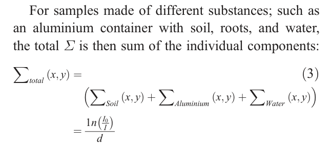
The component accounting for the water in the system, Σwater, comprises from both soil and root water. If the root thickness and the soil water content are known, the total Σ of water can be calculated from the water residing in the soil and water in the roots:
体系中代表水分的分量Σwater包括土壤水和根系水两部分。若已知根系粗度和土壤含水量,则可根据土壤中的水和根系中的水计算水的总Σ:

where d is the container thickness filled with soil and x is the root thickness.
式中,d为装填土壤的容器厚度,x为根系粗度。
We used the same method as Menon et al. (2007) for detecting root thickness and segmentation. All other calculations above apart from the neutron scattering correction were carried out using Matlab.
根系粗度检测和图像分割采用与Menon et al.(2007)相同的方法。除中子散射校正外,上述其他计算均使用Matlab完成。
9Results结果
10The analysis of a neutron radiograph中子成像图像分析
therefore a mean transmission value of 0.99. The contrast between the roots and the soil was due to their different θ.
因此平均透射率为0.99。根系与土壤之间的对比度源于二者θ的差异。
The effect of plant-growth medium on root contrast neutrons in the organic soil and the mine tailings can be attributed to their high concentrations in hydrogen and iron, respectively.
植物生长介质对根系对比度的影响 有机土壤和尾矿对中子的(高衰减)可分别归因于其高浓度的氢和铁。
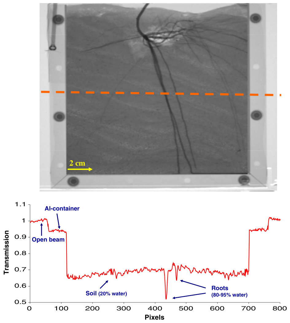
Figure 2 shows a NR image of a root system grown in an aluminium container filled with soil together with the transmission values along a cross-section through the image. There was a clear contrast between the soil and the roots, although the signal from the soil was heterogeneous due to the differences in the soil moisture content and the soil-filling patterns. Normalizing the image set a mean transmission value of 1 for the open beam area, while the sample area had different transmission based on its Σ. The aluminium frame of the container with a thickness of 16 mm absorbed only 5% of the incident neutrons resulting in a transmission value of 0.95. The aluminium sidewalls of the container had a thickness of 3 mm and图2为生长于装土铝制容器中的根系的NR图像,以及沿图像某一横截面的透射率值。土壤与根系之间对比明显,但由于土壤含水量和土壤装填方式的差异,土壤信号呈非均质性。图像归一化后,开束区域的平均透射率为1,而样品区域则依其Σ呈现不同的透射率。容器铝框厚度为16 mm,仅吸收了5%的入射中子,透射率为0.95。容器铝侧壁厚度为3 mm,
Fig. 2 Neutron radiograph of Berkheya coddii roots in an aluminium container filled with soil. The graph shows the transmission values of the pixels along the cross-section in the image图2 装土铝制容器中Berkheya coddii根系的中子成像图像。曲线图显示图像中沿横截面各像素的透射率值
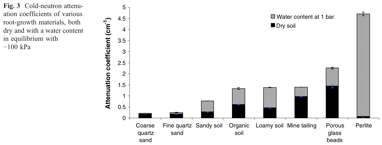
For most plants, the optimal soil moisture for growth is between field capacity (ca −33 kPa) and a water potential of −100 kPa (Allen et al. 1998). Reducing the soil θ improves contrast. Therefore, the optimal soil moisture for imaging roots by NR is at a soil water potential at the dry end of this range (−100 kPa). Plotting the Σ of the plant growth media containing their corresponding θ at −100 kPa changed the trend completely (Fig. 3). While dry perlite had the lowest Σ, perlite had the highest θ (Table 1) and therefore also the highest Σ at −100 kPa.
对于大多数植物,最适生长的土壤水分介于田间持水量(约−33 kPa)与水势−100 kPa之间(Allen et al. 1998)。降低土壤θ可提高对比度。因此,NR根系成像的最佳土壤水分为该范围干端的土壤水势(−100 kPa)。绘制植物生长介质在−100 kPa下含有相应θ时的Σ后,趋势完全改变(图3)。干燥珍珠岩的Σ最低,但珍珠岩的θ最高(表1),因此在−100 kPa下其Σ也最高。
11Relationship between the Σ and θΣ与θ的关系
lower sensitivity than the one for cold neutrons. Therefore we used the cold-neutron calibration curve for the rest of the analysis.
较冷中子的灵敏度更低。因此,后续分析均采用冷中子校准曲线。
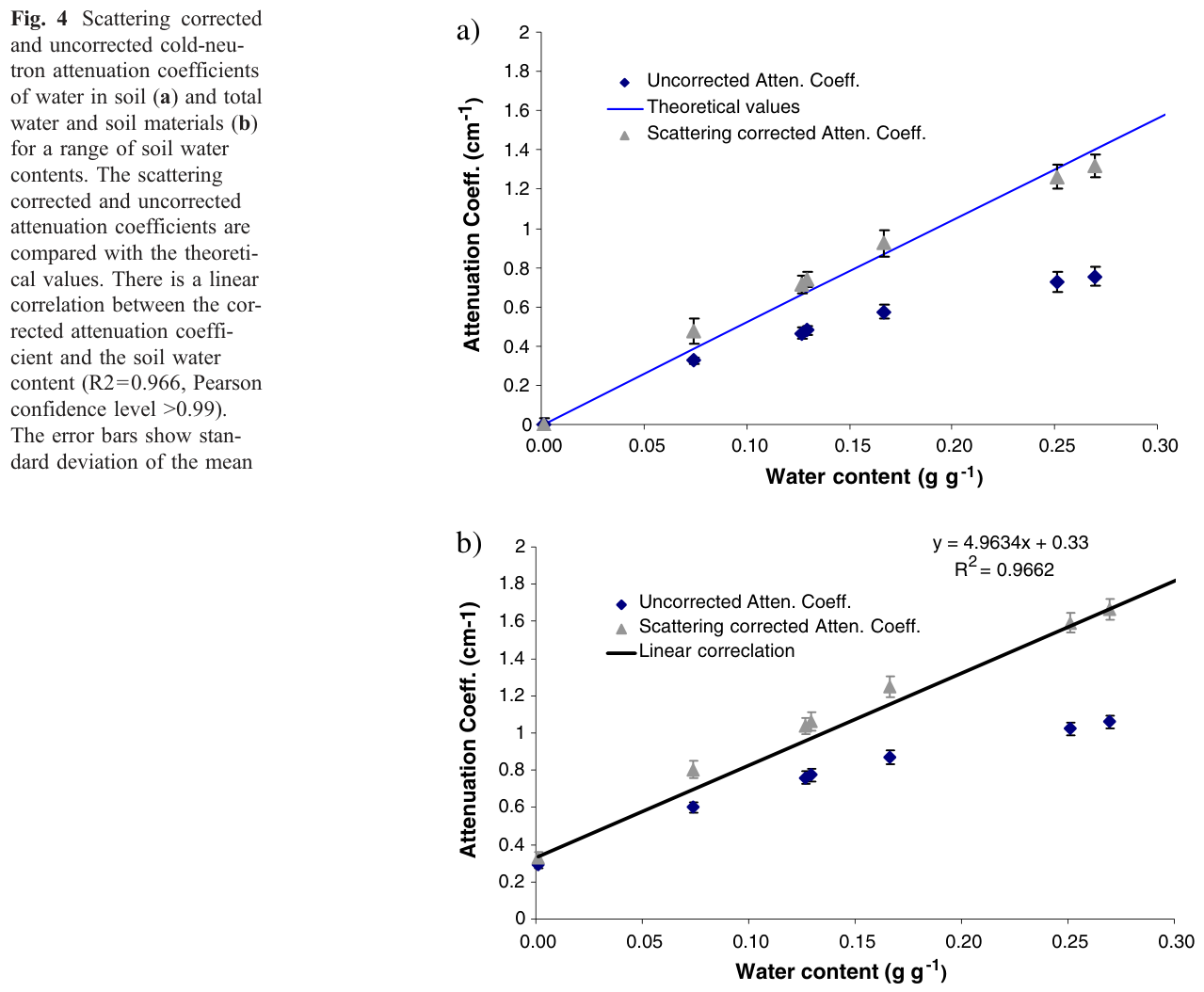
The total cold-neutron Σ (sum of soil and soil water Σ) followed the same trend as the neutron Σ of water (Fig. 4b). The intercept was equal to the Σ of the dry soil (0.33 cm−1). The relationship between the scattering-corrected total neutron Σ and soil θ was linear (R2=0.966, Pearson confidence level >0.99).
冷中子总Σ(土壤Σ与土壤水Σ之和)与水的中子Σ呈相同趋势(图4b)。截距等于干土的Σ(0.33 cm−1)。经散射校正的中子总Σ与土壤θ之间呈线性关系(R2=0.966,Pearson置信水平 >0.99)。
12Root detection根系检测
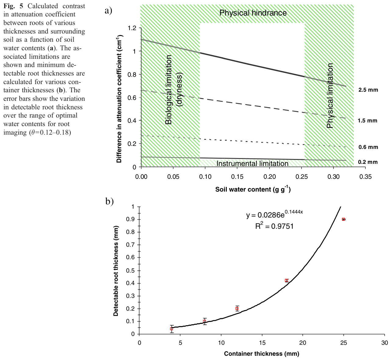
The ease of root detection in neutron radiographs increases in proportion to their θ and thickness. Coarser roots absorb or scatter more neutrons passing through the sample than fine roots. Therefore they create a better contrast with the surrounding soil. Figure 5a shows the contrast between roots and the surrounding soil (difference in Σ) for different root diameters over a range of soil θ. These calculations are based on extracted attenuation coefficient of soil in different water contents and equations 3 and 4 assuming a container thickness of 12 mm and an average root θ of 85%. Contrast increased with increasing root diameter and also with decreasing soil water content for each root diameter. The minimum diameter for root detection using our experimental setup was 0.2 mm. The pixel size of 110–170 μm and background noise arising from soil heterogeneity prevented the detection of finer roots. Roots were detectable when there was at least a 10% difference in Σ to the surrounding soil. At lower differences in Σ, measurement errors and background noise from soil heterogeneity prevented quantification.
在中子成像图像中,根系检测的难易程度随根系θ和粗度的增加而成比例改善。粗根比细根吸收或散射更多穿过样品的中子,因而与周围土壤形成更好的对比。图5a显示了不同根系直径在一系列土壤θ下根系与周围土壤之间的对比度(Σ差值)。这些计算基于提取的不同含水量下土壤的衰减系数以及式3和式4,并假设容器厚度为12 mm、根系平均θ为85%。对于每一根系直径,对比度均随根系直径的增大以及土壤含水量的降低而增加。采用本实验装置时,可检测的最小根系直径为0.2 mm。110–170 μm的像素尺寸以及由土壤非均质性引起的背景噪声妨碍了对更细根系的检测。当根系与周围土壤的Σ差异至少为10%时,根系即可被检测到。Σ差异更低时,测量误差和由土壤非均质性引起的背景噪声使定量无法进行。
At a soil θ below 0.1, the roots started dehydrating, which reduced contrast and inhibited plant growth. The flux of transmitted neutrons decreased at a soil θ above 0.25 due to high neutron scattering by soil water. This negatively affected the scattering correction and reduced contrast.
当土壤θ低于0.1时,根系开始脱水,从而降低了对比度并抑制了植物生长。当土壤θ高于0.25时,由于土壤水对中子的强烈散射,透射中子通量下降。这对散射校正产生了不利影响,并降低了对比度。
Minimum detectable root diameter as a function of container thickness
可检测最小根系直径与容器厚度的关系
Our container had an inner width of 12 mm in the direction of the neutron beam in order to provide enough space for the roots to grow. Others have used root-growth boxes that varied in thickness from 6 mm to more than 30 mm (Furukawa et al. 1999; Kuchenbuch and Ingram 2002; Nakanishi et al.
为给根系生长提供足够的空间,本研究所用容器沿中子束方向的内宽为12 mm。其他研究者所用根箱厚度从6 mm到30 mm以上不等(Furukawa et al. 1999; Kuchenbuch and Ingram 2002; Nakanishi et al.
2003). Figure 5b shows how the minimum diameter of detectable roots depended on container thickness. This calculation was based on our results for the sandy soil using equations 3 and 4, assuming root θ of 85%, a soil θ of 0.12–0.18, a contrast threshold of 10% and no limitation in pixel size. With a container thickness of 4 mm, roots with a diameter of 0.04 mm could be resolved. The detectable root thickness increased exponentially with increasing the container width. With a container width of 25 mm, only roots thicker than 0.9 mm could be detected. The error bars show the variation in detectable root thickness over the range of optimal water contents for root imaging (θ=0.12–0.18). As the container thickness increased, the error bars became smaller which means that the influence of soil θ on root detection decreased.
2003)。图5b显示了可检测根系最小直径与容器厚度的关系。该计算基于本研究砂质土壤的结果,采用式3和式4,并假设根系θ为85%、土壤θ为0.12–0.18、对比度阈值为10%且像素尺寸不受限制。容器厚度为4 mm时,可分辨直径为0.04 mm的根系。可检测根系粗度随容器宽度的增加呈指数增长。容器宽度为25 mm时,仅能检测到粗于0.9 mm的根系。误差线表示在根系成像最佳含水量范围(θ=0.12–0.18)内可检测根系粗度的变化。随着容器厚度增加,误差线变小,表明土壤θ对根系检测的影响减弱。
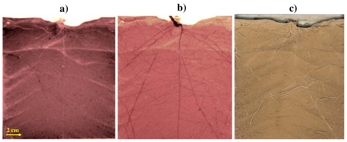
NR, X-ray radiographs and scan photos of the plant roots appeared darker than the surrounding soil in neutron radiograph due to their higher neutron attenuation coefficient than the soil. Not only main roots, but also lateral roots were also clearly visible. Figure 6c shows a scan of the soil surface after opening one side of the container. This method only showed roots on the surface and could not give a picture of roots enmeshed in the opaque soil.
NR、X射线成像与扫描照片 在中子成像图像中,植物根系由于中子衰减系数高于土壤而显得比周围土壤更暗。不仅主根,侧根也清晰可见。图6c为打开容器一侧后土壤表面的扫描图像。该方法只能显示表面的根系,无法呈现交织于不透明土壤中的根系。
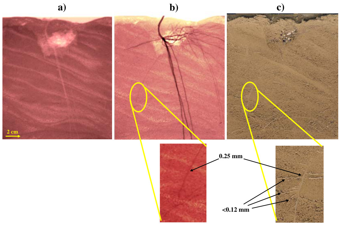
The X-Ray and cold-neutron images of Berkheya coddii (Fig. 7a and b) had similar characteristics as the respective images of Cicer arietinum. Because Berkheya coddii also produces fine roots (<2 mm in diameter) in addition to the coarse main roots, the scan of the opened side of the soil (Fig. 7c) revealed many roots that were not visible in the neutron radiograph. The enlargement in Fig. 7c shows roots with thickness of 0.12 mm that could not be seen in the neutron radiograph because it was below the rootdetection limit (0.2 mm). However, the root with thickness of 0.25 mm was clearly visible in both images.
Berkheya coddii的X射线和冷中子图像(图7a和b)与Cicer arietinum相应图像的特征相似。由于Berkheya coddii除粗大主根外还产生细根(直径<2 mm),打开一侧后的土壤扫描图像(图7c)显示出许多在中子成像图像中不可见的根系。图7c中的放大图显示了粗度为0.12 mm的根系,由于其低于根系检测限(0.2 mm),在中子成像图像中无法看到。然而,粗度为0.25 mm的根系在两幅图像中均清晰可见。
only the main roots and the roots on the surface respectively, the neutron image reveals both fine roots and roots within the soil
仅显示主根和表面的根系,而中子图像则同时显示了细根和土壤内部的根系
Lycopersicon esculentum produces a fibrous rooting system of which most of the roots are in the range of 0.2 mm and thinner. Comparing the scanned image (Fig. 8c) with the neutron radiograph (Fig. 8b) revealed many fine roots that were only visible in the scanned image. These roots were mostly located at the bottom part of the container. In the X-Ray radiograph (Fig. 8a) of this plant, no roots were visible except for the main root which had a thickness of more than 7 mm and was clearly visible.
Lycopersicon esculentum具有须根系,其大部分根系粗度在0.2 mm及以下。将扫描图像(图8c)与中子成像图像(图8b)比较,发现许多细根仅在扫描图像中可见。这些根系大多位于容器底部。在该植株的X射线成像图像(图8a)中,除粗度超过7 mm且清晰可见的主根外,未见其他根系。
roots than the other two techniques, but it fails to detect roots thinner than 0.2 mm, which are clearly visible in the scan image
根系多于其他两种技术,但其无法检测到粗度小于0.2 mm的根系,而这些根系在扫描图像中清晰可见
13Discussions讨论
Root-soil contrast and choice of plant-growth medium
根系-土壤对比度与植物生长介质的选择
While soil contains typically less than 50% water by mass (in our case 20%), the θ of roots usually is in the range of 80–95%. Therefore, roots are best seen in soils with a low θ, low intrinsic Σ (meaning low concentrations of high neutron-absorbing elements), and low heterogeneity.
土壤的含水量通常低于50%(质量比;本研究中为20%),而根系的θ通常在80–95%范围内。因此,在θ低、固有Σ低(即强中子吸收元素浓度低)且非均质性低的土壤中,根系最易被观察到。
The choice of the plant growth medium is of overriding importance for root quantification with NR. An ideal plant growth medium for neutron imaging should provide enough contrast with roots, i.e. the lowest possible Σ, while providing a satisfactory environment for plant growth. The sandy soil (Fig. 3) was the best medium since it had a relatively low Σ at both dry and wet conditions and still supported plant growth without water stress. Although fine and coarse quartz sands had lower Σ than the sandy soil, mechanical resistance to root penetration due to high bulk density can be a problem for root growth (Menon et al. 2007).
植物生长介质的选择对于利用NR定量根系至关重要。理想的中子成像植物生长介质应在为植物生长提供适宜环境的同时,与根系形成足够的对比,即Σ应尽可能低。砂质土壤(图3)是最佳介质,因为它在干、湿条件下均具有相对较低的Σ,且仍能支持植物在无水分胁迫的情况下生长。尽管细石英砂和粗石英砂的Σ低于砂质土壤,但其容重较高,对根系穿透的机械阻力可能成为根系生长的问题(Menon et al. 2007)。
14Correcting for scattering散射校正
Quantitative analysis of soil water and root distribution using NR requires scattering correction, especially when high concentrations of hydrogen are present in the system. Scattering had a large effect on radiographs taken within the range of soil θ that are optimum for root growth in most of the soils. Correcting for scattering improved the linearity between soil θ and the corresponding neutron attenuation coefficients (Fig. 4). Such linearity greatly enhances the reliability and sensitivity (up to 80%) of the calibration curve and simplifies the use of the calibration curve for relating water content to the attenuation coefficient which is crucial for quantitative analysis of roots and water distribution in soil by means of NR.
利用NR定量分析土壤水分和根系分布需要进行散射校正,尤其是当体系中存在高浓度氢时。对于大多数土壤,在最适根系生长的土壤θ范围内拍摄的成像图像受散射影响很大。散射校正改善了土壤θ与相应中子衰减系数之间的线性关系(图4)。这种线性关系极大地提高了校准曲线的可靠性和灵敏度(最高达80%),并简化了利用校准曲线将含水量与衰减系数相关联的过程,而这对于借助NR定量分析土壤中根系和水分分布至关重要。
15NR in relation to X-ray and scan photosNR与X射线成像及扫描照片的比较
NR revealed the details of the roots the most among the other two methods (Figs. 6, 7 and 8). X-ray radiographs had a weaker contrast between the roots and the soil than NR. Using microfocus X-Ray sources could improve the spatial resolution and the contrast between roots and the surrounding soil. Although our X-Ray imaging system had a large focal spot, (3 mm), it provided detailed information on the soil structure: textural layers created by the filling process and the cracks in the soil were clearly visible. Therefore, X-Ray and neutron images provide complementary information on the root-soil system. X-Ray radiography, which provides information on soil texture, could be used to enhance root contrast by revealing the position of non-root features in the rhizosphere.
与另外两种方法相比,NR对根系细节的揭示最为充分(图6、图7和图8)。X射线成像图像中根系与土壤之间的对比度弱于NR。使用微焦点X射线源可提高空间分辨率以及根系与周围土壤之间的对比度。尽管本研究的X射线成像系统焦斑较大(3 mm),但其提供了详细的土壤结构信息:装填过程形成的质地分层以及土壤中的裂隙均清晰可见。因此,X射线图像与中子图像可提供关于根系-土壤体系的互补信息。X射线成像可提供土壤质地信息,可通过揭示根际中非根系特征的位置来增强根系对比度。
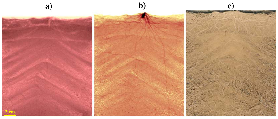
especially in the bottom of the container (which is clearly visible in the scan image) although it reveals the thicker roots in the upper part of the container
尤其是容器底部的细根(在扫描图像中清晰可见),但显示了容器上部较粗的根系
Scan images provided details of the roots only on the soil surface. Scan images could be useful to estimate the proportion of roots with a diameter of <2 mm, which were not detectable using NR. These very fine roots are mostly lost when roots are physically separated from soil.
扫描图像仅提供了土壤表面根系的细节。扫描图像可用于估算NR无法检测的直径<2 mm根系所占比例。当根系从土壤中物理分离时,这些极细的根系大多会损失。
Advantages and limitations of NR for the visualisation of plant roots
NR用于植物根系可视化的优势与局限性
With respect to quantitative root system analysis, NR application is currently still limited at high soil θ due to high neutron scattering, and at low soil θ, due to root dehydration (Fig. 5a). Improvements in instrumentation, in particular, higher resolution detector systems, will permit the visualisation of roots with a diameter of <0.2 mm. Nevertheless, NR currently can image roots with a thickness of ≥0.2 mm and in a range of soil θ (0.1–0.25) that is well suited for plant growth. Using thermal neutrons with higher energy and, therefore higher penetration, permits the visualisation of root growth at soil θ>0.25 (data not shown), although the contrast is reduced.
就根系的定量分析而言,NR的应用目前仍受到限制:在土壤θ较高时受中子强烈散射的限制,在土壤θ较低时则受根系脱水的限制(图5a)。仪器的改进,尤其是更高分辨率的探测系统,将使直径<0.2 mm的根系得以显像。尽管如此,NR目前已能在非常适合植物生长的土壤θ范围(0.1–0.25)内对粗度≥0.2 mm的根系进行成像。使用能量更高、因而穿透力更强的热中子,可在土壤θ>0.25时实现根系生长的可视化(数据未列出),但对比度会降低。
Acknowledgements This study was funded by the Swiss National Science Foundation. Dr. Héctor Conesa had a grant from Fundación Séneca of Comunidad Autónoma de Murcia (Spain).
致谢 本研究由瑞士国家科学基金会资助。Héctor Conesa博士获得了(西班牙)穆尔西亚自治区Fundación Séneca的资助。
16References参考文献
Allen RG, Pererira LS, Raes D, Smith M (1998) Crop evapotranspiration. Guidelines for computing crop water requirements. FAO, Rome
Bailey IF (2003) A review of sample environments in neutron scattering. Zeitschrift Fur Kristallographie 218:84–95
Bois JF, Couchat P (1983) Comparison of the effects of waterstress on the root systems of 2 cultivars of upland rice (Oryza-Sativa-L). Ann Bot 52:479–487
Bottomley PA, Rogers HH, Prior SA (1993) Nmr imaging of root water distribution in intact Vicia-Faba L plants in elevated atmospheric Co2. Plant Cell Environ 16:335–338
Costa C, Dwyer LM, Hamel C, Muamba DF, Wang XL, Nantais L, Smith DL (2001) Root contrast enhancement for measurement with optical scanner-based image analysis. Can J Bot-Revue Canadienne De Botanique 79:23–29
Doussan C, Pages L, Pierret A (2003) Soil exploration and resource acquisition by plant roots: an architectural and modelling point of view. Agronomie 23:419–431
Fitz W, Puschenreiter M, Schweiger P, Wenzel WW (2005) Novel tools for investigating rhizosphere processes. Abstracts of Papers of the American Chemical Society 230:U1789–U1790
Furukawa J, Nakanishi TM, Matsubayashi H (1999) Neutron radiography of a root growing in soil with vanadium. Nucl Instrum Methods Phys Res Sect A 424:116–121
Gregory PJ, Hutchison DJ, Read DB, Jenneson PM, Gilboy WB, Morton EJ (2003) Non-invasive imaging of roots with high resolution X-ray micro-tomography. Plant Soil 255:351–359
Hall LD, Amin MHG, Dougherty E, Sanda M, Votrubova J, Richards KS, Chorley RJ, Cislerova M (1997) MR properties of water in saturated soils and resulting loss of MRI signal in water content detection at 2 tesla. Geoderma 80:431–448
Hassanein RK (2006) Correction methods for the quantitative evaluation of thermal neutron tomography. ETH Zurich, Switzerland
Hassanein R, Lehmann E, Vontobel P (2005) Methods of scattering corrections for quantitative neutron radiography. Nucl Instrum Methods Phys Res Sect A 542:353–360
Heyes JA, Clark CJ (2003) Magnetic resonance imaging of water movement through asparagus. Funct Plant Biol 30:1089–1095
Hoagland DR, Arnon DI (1938) The water culture method for growing plants without soil. Calif Agric Exp Stn 347:1–39 Kasperl S, Vontobel P (2005) Application of an iterative artefact reduction method to neutron tomography. Nucl Instrum Methods Phys Res Sect A 542:392–398
Kuchenbuch RO, Ingram KT (2002) Image analysis for non-destructive and non-invasive quantification of root growth and soil water content in rhizotrons. Journal of Plant Nutrition and Soil Science-Z PflanzenernaÉhr Bodenkd 165:573–581
Kuhne G, Frei G, Lehmann E, Vontobel P (2005) CNR—the new beamline for cold neutron imaging at the Swiss spallation neutron source SINQ. Nucl Instrum Methods Phys Res Sect A 542:264–270
Lehmann E, Pleinert H, Williams T, Pralong C (1999) Application of new radiation detection techniques at the Paul Scherrer Institut, especially at the spallation neutron source. Nucl Instrum Methods Phys Res Sect A 424:158– 164
Majdi H (1996) Root sampling methods—Applications and limitations of the minirhizotron technique. Plant Soil 185:255–258
Matsushima U, Kawabata Y, Hino M, Geltenbort P, Nicolai BM (2005) Measurement of changes in water thickness in plant materials using very low-energy neutron radiography. Nucl Instrum Methods Phys Res Sect A 542:76–80
Menon M, Robinson B, Oswald SE, Kaestner A, Abbaspour KC, Lehmann E, Schulin R (2007) Visualization of root growth in heterogeneously contaminated soil using neutron radiography. Eur J Soil Sci 58:802–810
Moran CJ, Pierret A, Stevenson AW (2000) X-ray absorption and phase contrast imaging to study the interplay between plant roots and soil structure. Plant Soil 223:99–115
Naftel SJ, Martin RR, Sham TK, Macfie SM, Jones KW (2001) Micro-synchrotron X-ray fluorescence of cadmium-challenged corn roots. J Electron Spectrosc Relat Phenom 119:235–239
Nakaji T, Noguchi K, Oguma H (2008) Classification of rhizosphere components using visible-near infrared spectral images. Plant Soil 310:245–261
Nakanishi TM, Okuni Y, Furukawa J, Tanoi K, Yokota H, Ikeue N, Matsubayashi M, Uchida H, Tsiji A (2003) Water movement in a plant sample by neutron beam analysis as well as positron emission tracer imaging system. J Radioanal Nucl Chem 255:149–153
Nakanishi TM, Okuni Y, Hayashi Y, Nishiyama H (2005) Water gradient profiles at bean plant roots determined by neutron beam analysis. J Radioanal Nucl Chem 264:313–317
Oswald SE, Menon M, Robinson B, Carminati A, Vontobel P, Lehmann E, Schulin R (2008) Quantitative imaging of infiltration, root growth, and root water uptake via neutron radiography. Vadose Zone Journal 7:1035–1047
Pierret A (2008) Multi-spectral imaging of rhizobox systems: new perspectives for the observation and discrimination of rhizosphere components. Plant Soil 310:263–268
Pierret A, Doussan C, Garrigues E, Mc Kirby J (2003a) Observing plant roots in their environment: current
imaging options and specific contribution of two-dimensional approaches. Agronomie 23:471–479
Pierret A, Kirby M, Moran C (2003b) Simultaneous X-ray imaging of plant root growth and water uptake in thin-slab systems. Plant Soil 255:361–373
Pierret A, Moran CJ, Doussan C (2005) Conventional detection methodology is limiting our ability to understand the roles and functions of fine roots. New Phytol 166:967–980
Sutton RF, Tinus RW (1983) Root and root system terminology. For Sci Monogr 24:137
Tumlinson LG, Liu HY, Silk WK, Hopmans JW (2008) Thermal neutron computed tomography of soil water and plant roots. Soil Sci Soc Am J 72:1234–1242
Vogt KA, Vogt DJ, Bloomfield J (1998) Analysis of some direct and indirect methods for estimating root biomass and production of forests at an ecosystem level. Plant Soil 200:71–89
Waisel Y, Eshel A, Kafkafi Z (Eds.) (2002) Plant roots: the hidden half. Marcel Dekker, New York
Willatt ST, Struss RG (1979) Germination and early growth of plants studied using neutron radiography. Ann Bot 43:415–422
Willatt ST, Struss RG, Taylor HM (1978) Insitu root studies using neutron radiography. Agron J 70:581–586